. 2020 Mar;15(3):528-536.
doi: 10.4103/1673-5374.266060.

Ferrostatin-1 protects HT-22 cells from oxidative toxicity

Affiliations

Ferrostatin-1 protects HT-22 cells from oxidative toxicity

Jun Chu et al. Neural Regen Res. 2020 Mar.

Abstract

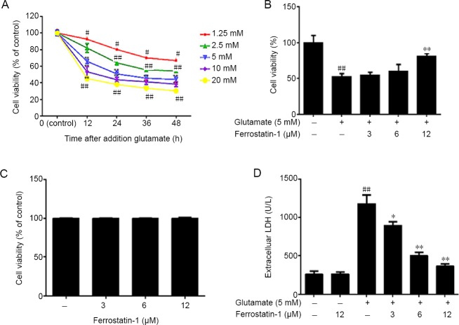
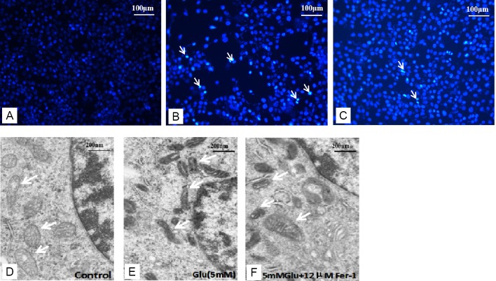
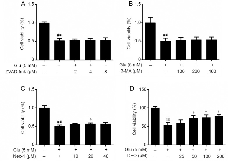
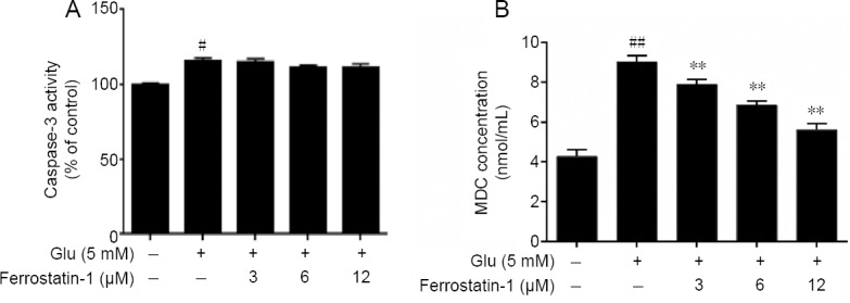
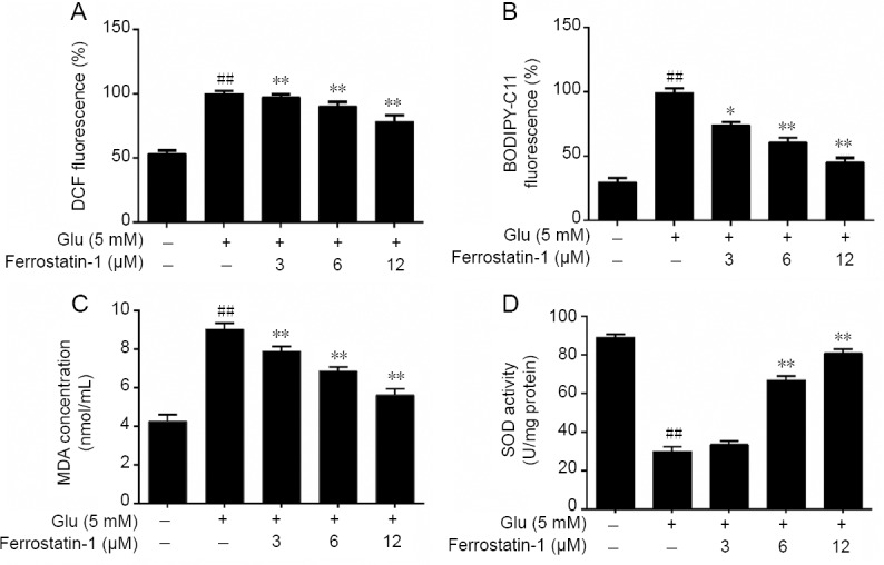
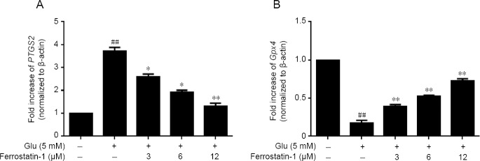
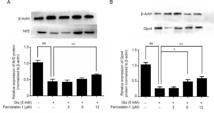
Ferroptosis is a type of programmed cell death dependent on iron. It is different from other forms of cell death such as apoptosis, classic necrosis and autophagy. Ferroptosis is involved in many neurodegenerative diseases. The role of ferroptosis in glutamate-induced neuronal toxicity is not fully understood. To test its toxicity, glutamate (1.25-20 mM) was applied to HT-22 cells for 12 to 48 hours. The optimal experimental conditions occurred at 12 hours after incubation with 5 mM glutamate. Cells were cultured with 3-12 μM ferrostatin-1, an inhibitor of ferroptosis, for 12 hours before exposure to glutamate. The cell viability was detected by 3-(4,5-dimethylthiazol-2-yl)-2,5-diphenyltetrazolium bromide assay. Autophagy was determined by monodansylcadaverine staining and apoptosis by caspase 3 activity. Damage to cell structures was observed under light and by transmission electron microscopy. The release of lactate dehydrogenase was detected by the commercial kit. Reactive oxygen species were measured by flow cytometry. Glutathione peroxidase activity, superoxide dismutase activity and malondialdehyde level were detected by the appropriate commercial kit. Prostaglandin peroxidase synthase 2 and glutathione peroxidase 4 gene expression was detected by real-time quantitative polymerase chain reaction. Glutathione peroxidase 4 and nuclear factor erythroid-derived-like 2 protein expression was detected by western blot analysis. Results showed that ferrostatin-1 can significantly counter the effects of glutamate on HT-22 cells, improving the survival rate, reducing the release of lactate dehydrogenase and reducing the damage to mitochondrial ultrastructure. However, it did not affect the caspase-3 expression and monodansylcadaverine-positive staining in glutamate-injured HT-22 cells. Ferrostatin-1 reduced the levels of reactive oxygen species and malondialdehyde and enhanced superoxide dismutase activity. It decreased gene expression of prostaglandin peroxidase synthase 2 and increased gene expression of glutathione peroxidase 4 and protein expressions of glutathione peroxidase 4 and nuclear factor (erythroid-derived)-like 2 in glutamate-injured HT-22 cells. Treatment of cultured cells with the apoptosis inhibitor Z-Val-Ala-Asp (OMe)-fluoromethyl ketone (2-8 μM), autophagy inhibitor 3-methyladenine (100-400 μM) or necrosis inhibitor necrostatin-1 (10-40 μM) had no effect on glutamate induced cell damage. However, the iron chelator deferoxamine mesylate salt inhibited glutamate induced cell death. Thus, the results suggested that ferroptosis is caused by glutamate-induced toxicity and that ferrostatin-1 protects HT-22 cells from glutamate-induced oxidative toxicity by inhibiting the oxidative stress.

Keywords: HT-22 cell; ferroptosis; ferrostatin-1; glutamate; glutathione peroxidase 4; oxidative toxicity; prostaglandin peroxidase synthase 2; reactive oxygen species.


Conflict of interest statement

None

Figures

Figure 1
Figure 1
Effect of ferrostatin-1 on glutamate-induced toxicity in HT-22 cells. (A) The decreased cell viability, measured by 3-(4,5-dimethylthiazol-2-yl)-2,5-diphenyltetrazolium bromide (MTT) assay, was induced by increasing doses of glutamate treatment. (B) The effect of ferrostatin-1 on the viability of HT-22 cells incubated with 5 mM glutamate, as measured by MTT assay. (C) The effect of ferrostatin-1 on the HT-22 cells. Viability assayed with MTT. (D) The effect of ferrostatin-1 on the lactate dehydrogenase (LDH) level in the HT-22 cells, with and without the glutamate treatment. Data are expressed as the mean ± SD. #P < 0.05, ##P < 0.01, vs. control group; *P < 0.05, **P < 0.01, vs. glutamate group (one-way analysis of variance followed by Tukey’s post-hoc test). Quantification data represented the mean of three experiments in duplicates. HT-22 cell: A mouse hippocampal neurons cell.
Figure 2
Figure 2
Effect of ferrostatin-1 on the morphological features of HT-22 cells. (A–C) 4′,6-Diamidino-2-phenylindole (DAPI) staining of HT-22 cells in (A) control, (B) Glu (5 mM), (C) Glu (5 mM) + Fer-1 (12 μM) groups. The white arrows indicate cell nucleus. (D–F) Transmission electron microscopy was used to observe the mitochondria. The white arrow indicates mitochondria. The mitochondria membrane density increased after the Glu treatment (E) compared with the control group (D) but the changes were smaller in the Glu + Fer-1 group (F). Scale bars: 100 μm in A–C, 200 μm in D–F. Fer-1: Ferrostatin-1; Glu: glutamate; HT-22 cell: a mouse hippocampal neurons cell.
Figure 3
Figure 3
Effects of other types of cell death inhibitor including ZVAD-fmk (A), 3-MA (B), Nec-1 (C) and DFO (D) on the cell viability of HT-22 cells treated with Glu as detected by MTT. Cell viability are expressed the optical density ratio to control group. Data are expressed as the mean ± SD. ##P < 0.01, vs. control group; *P < 0.05, **P < 0.01, vs. Glu group (one-way analysis of variance followed by Tukey’s post-hoc test). Quantification data represented the mean of three experiments in duplicates. 3-MA: 3-Methyladenine; DFO: deferoxamine mesylate salt; Glu: glutamate; HT-22 cell: a mouse hippocampal neurons cell; MTT: 3-(4,5-dimethylthiazol-2-yl)-2,5-diphenyltetrazolium bromide; Nec-1: necrostatin-1; ZVAD-fmk: Z-Val-Ala-Asp (OMe)-fluoromethyl ketone.
Figure 4
Figure 4
Effects of glutamate and ferrostatin-1 on apoptosis and autophagy measured as the activity of caspase-3 (A) and MDC (B) in HT-22 cells. Data are expressed as the mean ± SD. #P < 0.05, ##P < 0.01, vs. control group; **P < 0.01, vs. Glu group (one-way analysis of variance followed by Tukey’s post-hoc test). Quantification data represented the mean of three experiments in duplicates. Glu: Glutamate; HT-22 cell: a mouse hippocampal neurons cell; MDC: monodansylcadaverine.
Figure 5
Figure 5
Effects of ferrostatin-1 on glutamate-induced ROS and lipid peroxidation level in Glu-treated HT-22 cells. (A, B) The effect of ferrostatin-1 on cytosolic (A) and lipid (B) ROS level in Glu-treated HT-22 cells by flow cytometry. (C, D) The effect of ferrostatin-1 on MDA (C) and SOD (D) level in Glu-treated HT-22 cells. Data are expressed as the mean ± SD. ##P < 0.01, vs. control group; *P < 0.05, **P < 0.01, vs. Glu group (one-way analysis of variance followed by Tukey’s post-hoc test). Quantification data represent the mean of three experiments in duplicates. Glu: Glutamate; HT-22 cell: a mouse hippocampal neurons cell; MDA: malondialdehyde; ROS: reactive oxygen species; SOD: superoxide dismutase.
Figure 6
Figure 6
Effect of ferrostatin-1 on the expression of PTGS2 (A) and Gpx4 (B) after Glu treatment in HT-22 cells. The data present as the mean ± SD. ##P < 0.01, vs. control group; *P < 0.05, **P < 0.01, vs. Glu group (one-way analysis of variance followed by Tukey’s post-hoc test). Quantification data represent the mean of three experiments in duplicates. Glu: Glutamate; Gpx4: glutathione peroxidase 4; HT-22 cell: a mouse hippocampal neurons cell; PTGS2: prostaglandin peroxidase synthase 2.
Figure 7
Figure 7
Effect of ferrostatin-1 on the expression of Nrf2 (A) and Gpx4 (B) after Glu treatment in HT-22 cells. Western blot data represent as the mean ± SD. ##P < 0.01, vs. control group; *P < 0.05, **P < 0.01, vs. Glu group (one-way analysis of variance followed by Tukey’s post-hoc test). Quantification data represent the mean of three experiments in duplicates. Glu: Glutamate; Gpx4: glutathione peroxidase 4; HT-22 cell: a mouse hippocampal neurons cell; Nrf2: nuclear factor erythroid-derived-like 2.

References

    1. Baba Y, Higa JK, Shimada BK, Horiuchi KM, Suhara T, Kobayashi M, Woo JD, Aoyagi H, Marh KS, Kitaoka H, Matsui T. Protective effects of the mechanistic target of rapamycin against excess iron and ferroptosis in cardiomyocytes. Am J Physiol Heart Circ Physiol. 2018;314:H659–H668. - PMC - PubMed
    1. Bogdan AR, Miyazawa M, Hashimoto K, Tsuji Y. Regulators of iron homeostasis: new players in metabolism, cell death, and disease. Trends Biochem Sci. 2016;41:274–286. - PMC - PubMed
    1. Brigelius-Flohe R, Muller M, Lippmann D, Kipp AP. The yin and yang of nrf2-regulated selenoproteins in carcinogenesis. Int J Cell Biol. 2012;2012:486147. - PMC - PubMed
    1. Cao JY, Dixon SJ. Mechanisms of ferroptosis. Cell Mol Life Sci. 2016;73:2195–2209. - PMC - PubMed
    1. Chen L, Hambright WS, Na R, Ran Q. Ablation of the ferroptosis inhibitor glutathione peroxidase 4 in neurons results in rapid motor neuron degeneration and paralysis. J Biol Chem. 2015;290:28097–28106. - PMC - PubMed