

Disseminated Invasive Candidiasis in an Immunocompetent Host
Affiliations
- PMID: 31571811
- PMCID: PMC6752813
Item in Clipboard
Disseminated Invasive Candidiasis in an Immunocompetent Host
Fed Pract.
2019 Sep.
Abstract
Health care providers should consider a nonbacterial source as the causative agent for invasive candidiasis infection in immunocompetent patients.
Copyright © 2019 Frontline Medical Communications Inc., Parsippany, NJ, USA.
Conflict of interest statement
Author disclosures The authors report no actual or potential conflicts of interest with regard to this article.
Figures
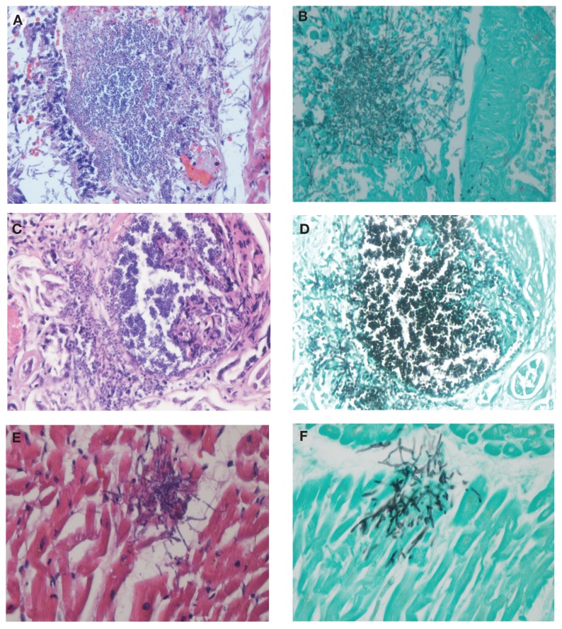
C albicans Colorization in Organs Due to Invasive Infection Abbreviations: C albicans, Candida albicans; H&E, hematoxylin and eosin; GMS, Grocott-Gömöri methenamine silver. Representative section of upper lobe of left lung H&E stain. (A) Acute bronchopneumonia with C albicans yeast and filaments surrounded with inflammatory reaction. (B) A corresponding GMS stain shows yeast and hyphae radiation in right lung tissues. (C) Huge inflammation in kidney matrix shows in a representative H&E stain section of right kidney. (D) GMS stain shows yeast predominant invasively multiplied C albicans inside a glomerulus. (E & F) Representative H&E and GMS section showing C albicans colonization in endocardium.
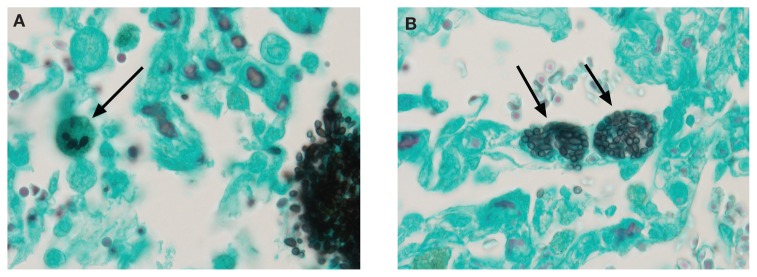
Candida albicans Colonization in the Lung (A) Histopathology of a dust cell with entrapped Candida albicans yeast (arrow); and (B) A lymphatic channel on alveolar septum with multiple yeast cells confirming dissemination of the organism systemically (arrows).
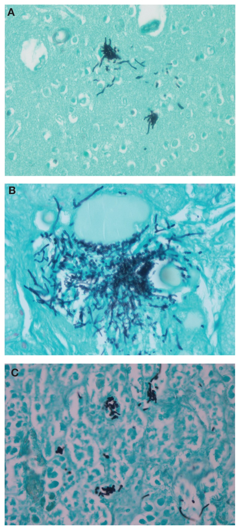
Candida albicans Colonization in Brain, Thyroid, and Adrenal Gland (A) Representative Grocott-Gömöri methenamine silver stain sections of brain; (B) thyroid; and (C) adrenal gland to show Candida albicans dissemination and colonization in other important organs.
Similar articles
-
Systemic candidiasis in an apparently immunocompetent dog.J Vet Diagn Invest. 2005 May;17(3):272-6. doi: 10.1177/104063870501700312. J Vet Diagn Invest. 2005. PMID: 15945387
-
Experimental Mouse Models of Disseminated Candida auris Infection.mSphere. 2019 Sep 4;4(5):e00339-19. doi: 10.1128/mSphere.00339-19. mSphere. 2019. PMID: 31484737 Free PMC article.
-
Modulation of neutrophil function in host defense against disseminated Candida albicans infection in mice.FEMS Immunol Med Microbiol. 1999 Dec;26(3-4):299-307. doi: 10.1111/j.1574-695X.1999.tb01402.x. FEMS Immunol Med Microbiol. 1999. PMID: 10575142 Review.
-
Invasive candidiasis during granulocytopenia.Recent Results Cancer Res. 1993;132:137-45. doi: 10.1007/978-3-642-84899-5_14. Recent Results Cancer Res. 1993. PMID: 8265854 Review.
-
Differential role of NK cells against Candida albicans infection in immunocompetent or immunocompromised mice.Eur J Immunol. 2014 Aug;44(8):2405-14. doi: 10.1002/eji.201343828. Epub 2014 May 27. Eur J Immunol. 2014. PMID: 24802993
Cited by
-
Septic shock due to candida and disseminated herpes simplex virus-1 (HSV1) after elective spinal surgery in an immunocompromised patient with chronic HSV1 infection.J Surg Case Rep. 2022 Jun 10;2022(6):rjac273. doi: 10.1093/jscr/rjac273. eCollection 2022 Jun. J Surg Case Rep. 2022. PMID: 35702262 Free PMC article.
-
Deep Cutaneous Candidiasis With Costal Osteomyelitis Following Pectoralis Major Myocutaneous Flap Reconstruction: A Case Report.Cureus. 2025 Jan 29;17(1):e78210. doi: 10.7759/cureus.78210. eCollection 2025 Jan. Cureus. 2025. PMID: 40027028 Free PMC article.
References
-
- Kullberg BJ, Arendrup MC. Invasive candidiasis. N Engl J Med. 2015;373(15):1445–1456. - PubMed
-
- Ericson EL, Klingspor L, Ullberg M, Ozenci V. Clinical comparison of the Bactec Mycosis IC/F, BacT/Alert FA, and BacT/Alert FN blood culture vials for the detection of candidemia. Diagn Microbiol Infect Dis. 2012;73(2):153–156. - PubMed
-
- Baum GL. The significance of Candida albicans in human sputum. N Engl J Med. 1960;263:70–73. - PubMed
LinkOut - more resources
Full Text Sources