

Correlation between laser speckle flowgraphy and optical coherence tomography angiography measurements in normal and glaucomatous eyes
- PMID: 31571818
- PMCID: PMC6750712
- DOI: 10.2147/OPTH.S213031
Correlation between laser speckle flowgraphy and optical coherence tomography angiography measurements in normal and glaucomatous eyes
Abstract
Purpose: To investigate the relationship between laser speckle flowgraphy (LSFG) and optical coherence tomography angiography (OCTA) measurements of the peripapillary retina and optic nerve head (ONH) in normal eyes and eyes with primary open-angle glaucoma (POAG).
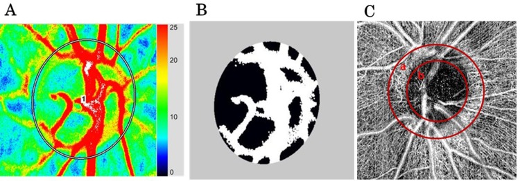
Patients and methods: One eye from each of 46 normal subjects and mild and moderate/advanced POAG patients were included. ONH blood flow acquired by LSFG, circumpapillary vessel density (cpVD, a 250 μm-wide elliptical annulus around the optic disc), and intra-papillary vessel density (ipVD, a 1.5×1.5 mm scan field) acquired by OCTA were measured. Their values were compared among normal controls and patients at each stage of glaucoma using one-way ANOVA, and the correlation between measurements obtained by the two methods was examined by univariate regression analysis.
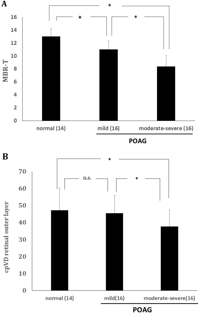
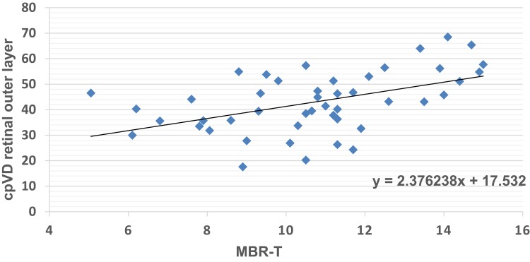
Results: ONH tissue blood flow, tissue mean blur rate (MBR-T), and cpVD in the outer layer of the retina significantly decreased with the progression of glaucoma stage, although the latter showed no significant difference between normal subjects and mild-stage glaucoma patients. MBR-T was significantly correlated with cpVD, but not with ipVD, in the retinal outer layer.
Conclusion: A correlation was found only between MBR-T and cpVD in the retinal outer layer. A difference in MBR-T, but not in cpVD, was detected between normal controls and mild glaucoma patients.
Keywords: blood flow; laser speckle flowgraphy; optic nerve head; optical coherent tomography angiography.
© 2019 Kohmoto et al.
Conflict of interest statement
The authors report no conflicts of interest in this work.
Figures
Similar articles
-
Ocular microcirculation measurement with laser speckle flowgraphy and optical coherence tomography angiography in glaucoma.Acta Ophthalmol. 2018 Jun;96(4):e485-e492. doi: 10.1111/aos.13639. Epub 2018 Mar 24. Acta Ophthalmol. 2018. PMID: 29575676
-
Correlation between structure/function and optic disc microcirculation in myopic glaucoma, measured with laser speckle flowgraphy.BMC Ophthalmol. 2014 Sep 24;14:113. doi: 10.1186/1471-2415-14-113. BMC Ophthalmol. 2014. PMID: 25252729 Free PMC article.
-
Relationship between laser speckle flowgraphy and optical coherence tomography angiography measurements of ocular microcirculation.Graefes Arch Clin Exp Ophthalmol. 2017 Aug;255(8):1633-1642. doi: 10.1007/s00417-017-3627-8. Epub 2017 May 1. Graefes Arch Clin Exp Ophthalmol. 2017. PMID: 28462456
-
[Aiming for zero blindness].Nippon Ganka Gakkai Zasshi. 2015 Mar;119(3):168-93; discussion 194. Nippon Ganka Gakkai Zasshi. 2015. PMID: 25854109 Review. Japanese.
-
[Nonarteritic ischemic optic neuropathy animal model and its treatment applications].Nippon Ganka Gakkai Zasshi. 2014 Apr;118(4):331-61. Nippon Ganka Gakkai Zasshi. 2014. PMID: 24864434 Review. Japanese.
Cited by
-
Change of intraocular blood flow during treatment for thyroid eye disease.Taiwan J Ophthalmol. 2022 Feb 28;12(1):97-100. doi: 10.4103/tjo.tjo_2_22. eCollection 2022 Jan-Mar. Taiwan J Ophthalmol. 2022. PMID: 35399966 Free PMC article.
-
Ocular blood flow as a clinical observation: Value, limitations and data analysis.Prog Retin Eye Res. 2020 Jan 24:100841. doi: 10.1016/j.preteyeres.2020.100841. Online ahead of print. Prog Retin Eye Res. 2020. PMID: 31987983 Free PMC article.
-
A New Approach to Retinal Oxygen Extraction Measurement Based on Laser Speckle Flowgraphy and Retinal Oximetry.Transl Vis Sci Technol. 2024 Dec 2;13(12):12. doi: 10.1167/tvst.13.12.12. Transl Vis Sci Technol. 2024. PMID: 39661379 Free PMC article. Clinical Trial.
-
Correlation Between Laser Speckle Flowgraphy and OCT-Derived Retinal and Choroidal Metrics in Healthy Human Eye.Transl Vis Sci Technol. 2022 Jun 1;11(6):15. doi: 10.1167/tvst.11.6.15. Transl Vis Sci Technol. 2022. PMID: 35704328 Free PMC article.
-
Ocular blood flow evaluation by laser speckle flowgraphy in pediatric patients with anisometropia.Front Public Health. 2023 Feb 27;11:1093686. doi: 10.3389/fpubh.2023.1093686. eCollection 2023. Front Public Health. 2023. PMID: 36923046 Free PMC article.
References
LinkOut - more resources
Full Text Sources
