. 2019 Sep 11:15:2629-2638.
doi: 10.2147/NDT.S220743. eCollection 2019.

Disrupted Brain Entropy And Functional Connectivity Patterns Of Thalamic Subregions In Major Depressive Disorder

Affiliations

Disrupted Brain Entropy And Functional Connectivity Patterns Of Thalamic Subregions In Major Depressive Disorder

Shao-Wei Xue et al. Neuropsychiatr Dis Treat. .

Abstract

Purpose: Entropy analysis of resting-state functional magnetic resonance imaging (R-fMRI) has recently been adopted to characterize brain temporal dynamics in some neuropsychological or psychiatric diseases. Thalamus-related dysfunction might be a potential trait marker of major depressive disorder (MDD), but the abnormal changes in the thalamus based on R-fMRI are still unclear from the perspective of brain temporal dynamics. The aim of this study was to identify local entropy changes and subregional connectivity patterns of the thalamus in MDD patients.

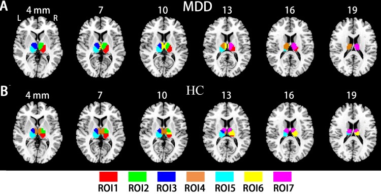
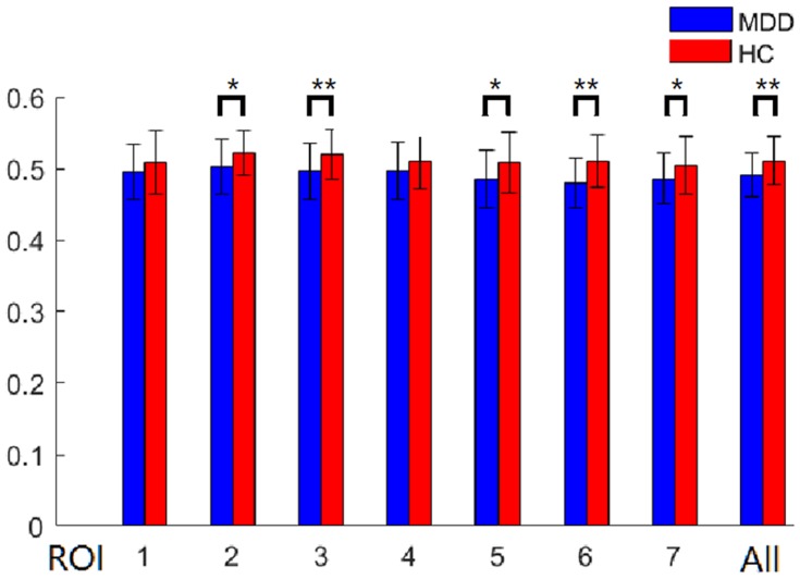
Patients and methods: We measured the sample entropy of the R-fMRI data from 46 MDD patients and 32 matched healthy controls. We employed the Louvain method for the module detection algorithm to automatically identify a functional parcellation of the thalamus and then examined the whole-brain subregional connectivity patterns.

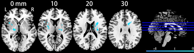
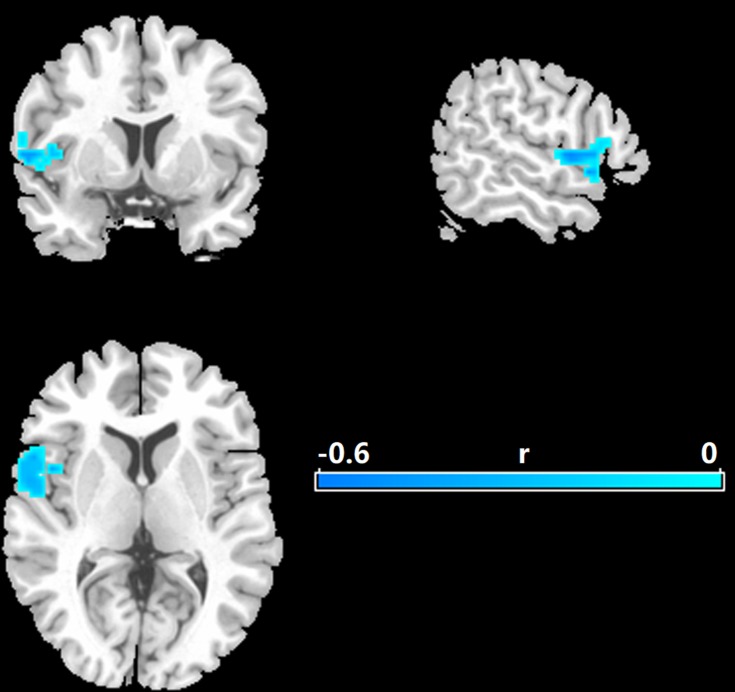
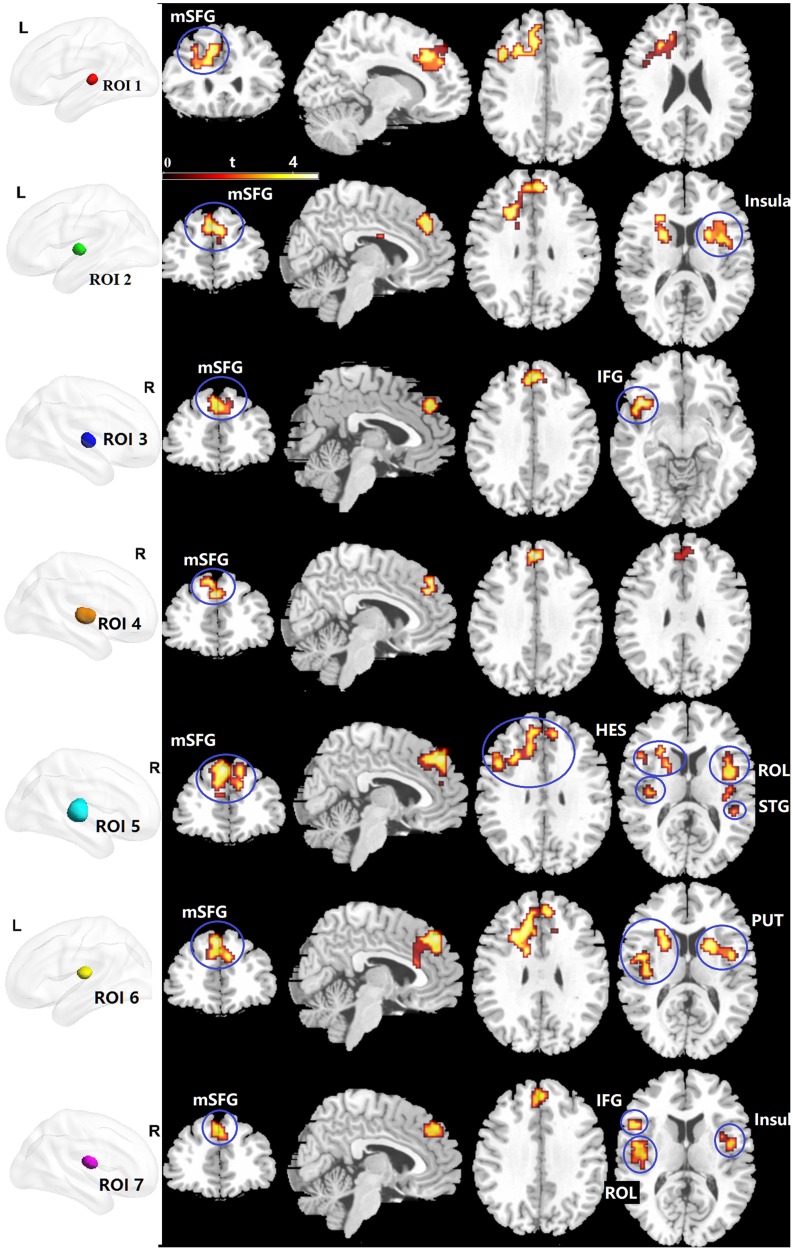
Results: The results indicated that the MDD patients had decreased entropy in the bilateral thalami compared with healthy controls. Increased functional connectivity between the thalamic subregions and the medial part of the superior frontal gyrus (mSFG) was found in MDD patients.

Conclusion: This study showed new evidence about sample entropy changes in MDD patients. The functional connectivity alterations that were widely distributed across almost all the thalamic subregions with the mSFG in MDD suggest a general involvement independent of the location and function of the subregions.

Keywords: entropy; functional connectivity; major depressive disorder; superior frontal gyrus; thalamus.


Conflict of interest statement

The authors report no conflicts of interest in relation to this work.

Figures

Figure 1
Figure 1
Clusters showing brain entropy differences in MDD patients compared with HC. Significance level was defined at voxel p < 0.005, cluster p < 0.05, GRF corrected. L refers to the left side of the images in the coronal, and horizontal sections correspond to the left side of the brain.
Figure 2
Figure 2
Correlations between entropy and the Hamilton rating scale for MDD. Significance level was defined at voxel p < 0.005, cluster p < 0.05, GRF corrected. L refers to the left side of the images in the coronal, and horizontal sections correspond to the left side of the brain.
Figure 3
Figure 3
The thalamic subregions. (A) Automated detection of seven thalamic subregions in the HC group. (B) Automated detection of seven thalamic subregions in the MDD group.
Figure 4
Figure 4
Between-group differences of subregional mean entropy. All, bilateral whole thalamus. *p < 0.05; ** P < 0.01. Abbreviations: ROI, region of interest; BE, brain entropy.
Figure 5
Figure 5
Clusters showing between-group RSFC differences of the thalamic subregions. Significance level was defined at voxel p < 0.005, cluster p < 0.05, GRF corrected. R refers to the left side of the image in the coronal and horizontal sections corresponding to the left side of the brain. Abbreviations: ROI, regions of interest; mSFG, the medial part of the superior frontal gyrus; IFG, inferior orbital frontal gyrus; HES, Heschl; ROL, Rolandic operculum; STG, superior temporal gyrus; PUT, putamen; L, left side.

Similar articles

Cited by

References

    1. Richards CS, O’Hara MW. The Oxford Handbook of Depression and Comorbidity. Oxford University Press; 2014:254.
    1. Kessler RC, Berglund P, Demler O, Jin R, Merikangas KR, Walters EE. Lifetime prevalence and age-of-onset distributions of DSM-IV disorders in the National Comorbidity Survey Replication. Arch Gen Psychiatry. 2005;62:593–602. doi:10.1001/archpsyc.62.6.593 - DOI - PubMed
    1. Belzung C, Willner P, Philippot P. Depression: from psychopathology to pathophysiology. Curr Opin Neurobiol. 2015;30:24–30. doi:10.1016/j.conb.2014.08.013 - DOI - PubMed
    1. Graham J, Salimi-Khorshidi G, Hagan C, et al. Meta-analytic evidence for neuroimaging models of depression: state or trait? J Affect Disord. 2013;151:423–431. doi:10.1016/j.jad.2013.07.002 - DOI - PubMed
    1. Gong Q, He Y. Depression, neuroimaging and connectomics: a selective overview. Biol Psychiatry. 2015;77:223–235. doi:10.1016/j.biopsych.2014.08.009 - DOI - PubMed