. 2019 Sep 16:15:2671-2680.
doi: 10.2147/NDT.S215263. eCollection 2019.

Galuteolin attenuates cerebral ischemia/reperfusion injury in rats via anti-apoptotic, anti-oxidant, and anti-inflammatory mechanisms

Affiliations

Galuteolin attenuates cerebral ischemia/reperfusion injury in rats via anti-apoptotic, anti-oxidant, and anti-inflammatory mechanisms

Xue Cheng et al. Neuropsychiatr Dis Treat. .

Abstract

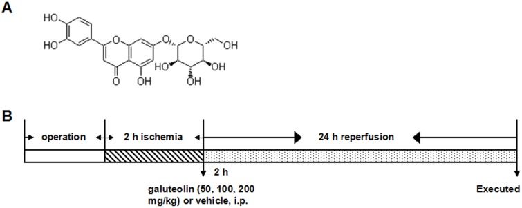
Purpose: Galuteolin is a substance extracted and purified from honeysuckle. The purpose of this study was to explore the protective effect of galuteolin on cerebral ischemia-reperfusion injury (CIRI) and reveal its potential molecular mechanism from the perspectives of anti-apoptosis, anti-oxidation, and anti-inflammation.

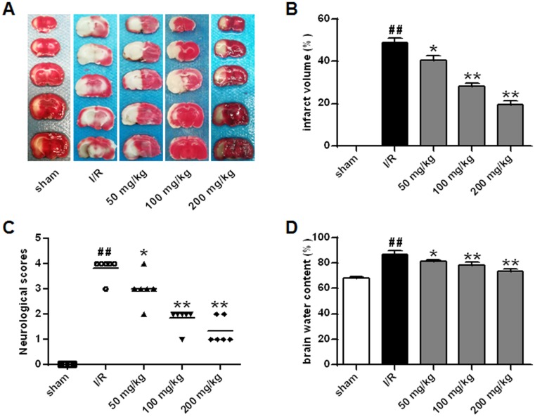
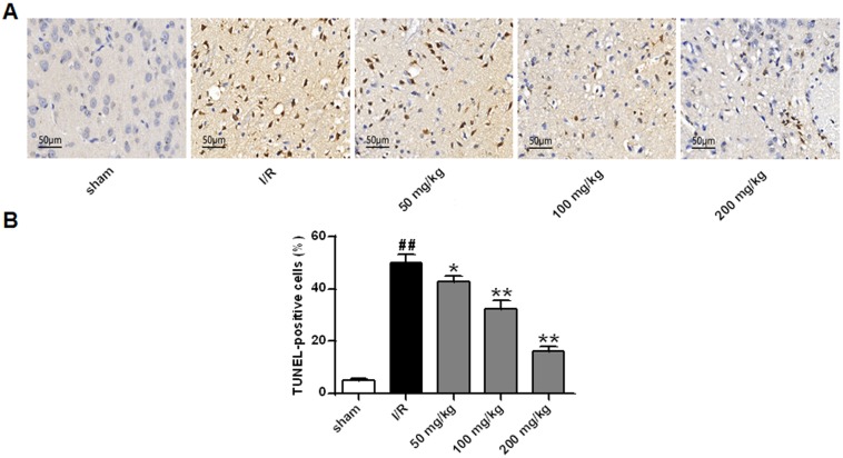
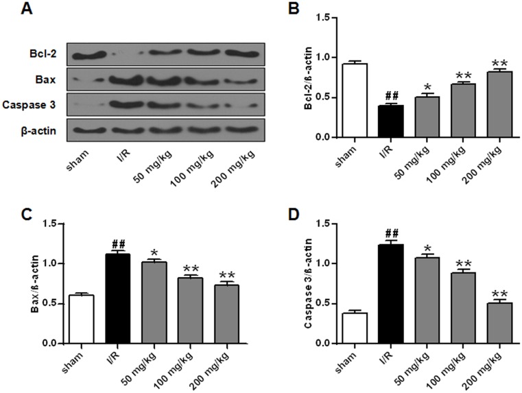
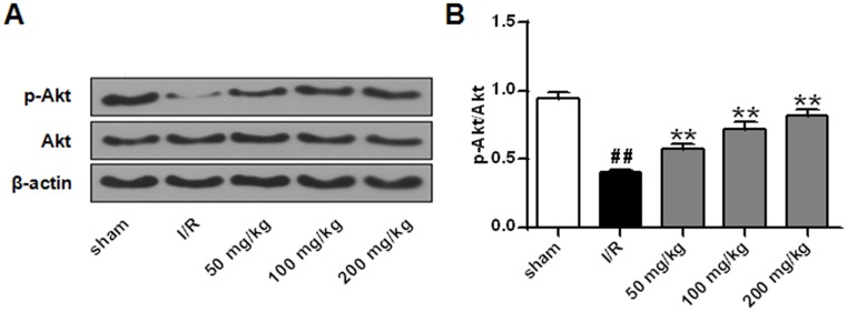
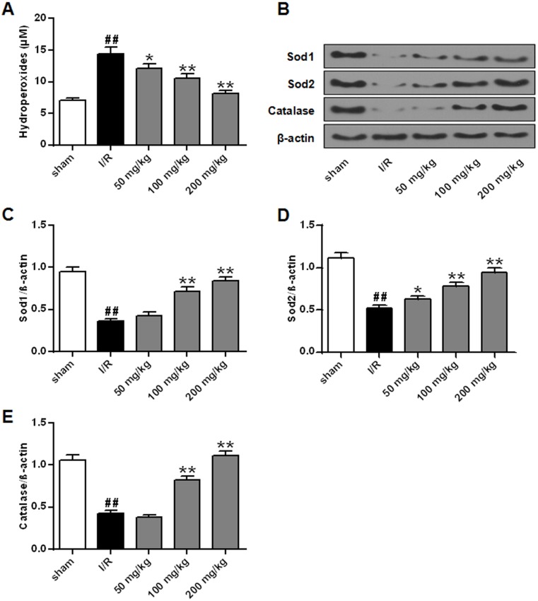
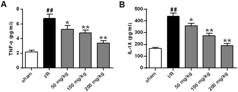
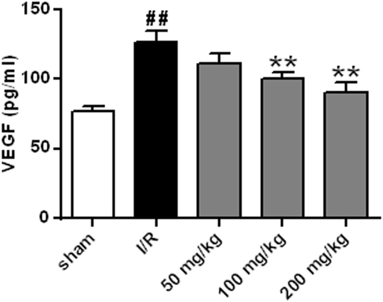
Materials and methods: One hundred and fifty rats were randomly divided into five groups: sham group, ischemia-reperfusion (I/R) group, 50 mg/kg galuteolin group, 100 mg/kg galuteolin group, and 200 mg/kg galuteolin group. Middle cerebral artery occlusion (MCAO) was used to establish a rat CIRI model, different doses of galtenolin were intraperitoneal injected 2 hrs after ischemia, and then reperfusion was performed for 24 hrs. Neurological function and cerebral water content were determined, and cerebral infarct volume was evaluated by TTC staining. TUNEL staining was used to detect the apoptosis of nerve cells. Western Blot was used to detect the expressions of Akt, p-Akt, Sod1, Sod2, catalase, caspase-3, Bcl-2, and Bax. Lipid hydrogen peroxide (LPO) was determined by kit assay. The contents of vascular endothelial growth factor (VEGF) and pro-inflammatory cytokines IL-1β and TNF-α were determined by ELISA.

Results: The results showed that galuteolin could significantly reduce the cerebral infarction volume, neurologic score, and cerebral water content in a dose-dependent manner. In addition, galuteolin obviously reduced the apoptosis rate of nerve cells and the expression levels of caspase-3 and Bax, meanwhile up-regulated the expression levels of p-Akt and Bcl-2. Furthermore, galuteolin apparently inhibited the levels of LPO, Sod1, Sod2, and catalase in the cerebral infarction tissues. Moreover, galuteolin also significantly reduced the levels of pro-inflammatory factors IL-1β and TNF-α in the cerebral infarction tissues. Finally, Galuteolin markedly inhibited the expression of VEGF in cerebral infarction tissues.

Conclusion: Galuteolin exerts neuroprotective effects against CIRI by inhibiting apoptosis, oxidation, and inflammation.

Keywords: apoptosis; galuteolin; inflammation; ischemia; oxidation; reperfusion.


Conflict of interest statement

The authors report no conflicts of interest in this work.

Figures

Figure 1
Figure 1
The chemical structure of galuteolin and the experimental process. (A) Schematic diagram of the chemical structure of galuteolin (C21H20O11, molecular weight: 448.38). (B) Flow chart of the establishment of rat MCAO/R model.
Figure 2
Figure 2
Effects of galuteolin on cerebral infarction volume, neural behavior, and cerebral water content after CIRI in rats. Effects of galuteolin on cerebral infarction volume (A, B), neural behavior, (C) and cerebral water content, and (D) after MCAO/R injury in rats. Notes: n=6, compared with the I/R group p, *P<0.05, **P<0.01; compared with the sham group, ##P<0.01.
Figure 3
Figure 3
Detection of the effect of galuteolin on neuronal apoptosis by TUNEL staining. (A) Images of TUNEL staining in rat brain tissue from each group. (B) TUNEL positive cells in the rat brain tissue of each group. Notes: n=6, compared with the I/R group p, *P<0.05, **P<0.01; compared with the sham group, ## P<0.01.
Figure 4
Figure 4
Effects of galuteolin on apoptosis-related proteins in rat cerebral infarction tissues. (A) Determination of the protein expressions of Bcl-2, Bax, and caspase-3 in cerebral infarction tissues of rats by Western Blot. (B) Histogram of BCL-2 protein expression. (C) Histogram of protein expression of Bax. (D) Histogram of caspase-3 protein expression. The β-actin as a standard internal reference. Notes: n=6, compared with the I/R group p, *P<0.05, **P<0.01; compared with the sham group, ## P<0.01.
Figure 5
Figure 5
Effects of galuteolin on Akt phosphorylation in rat cerebral infarction tissues. (A) Determination of the expressions of p-Akt and Akt in rat cerebral infarction by Western Blot. (B) Histogram of p-AktAkt protein expressions. The β-actin as a standard internal reference. Notes: n=6, compared with the I/R group, **P<0.01; compared with the sham group, ## P<0.01.
Figure 6
Figure 6
Effects of galuteolin on oxidative stress in cerebral infarction of rats. (A) Determination of the level of LPO in rat brain tissue by LPO kit. (B) Determination of the expression levels of Sod1, Sod2, and catalase by Western Blot. (C) Histogram of Sod1 protein expression. (D) Histogram of Sod2 protein expression. (E) Histogram of catalase protein expression. The β-actin as a standard internal reference. Notes: n=6, compared with the I/R group p, *P<0.05, **P<0.01; compared with the sham group, ## P<0.01.
Figure 7
Figure 7
Effects of galuteolin on inflammatory responses in rat cerebral infarction. (A) Determination of the secretion of pro-inflammatory cytokine TNF-α in cerebral infarction by ELISA. (B) Determination of the secretion of pro-inflammatory cytokine IL-1β in cerebral infarction by ELISA. Notes: n=6, compared with the I/R group p, *P<0.05, **P<0.01; compared with the sham group, ## P<0.01.
Figure 8
Figure 8
Effect of galuteolin on VEGF level in cerebral infarction tissue of rats. Notes: n=6, compared with the I/R group p, **P<0.01; compared with the sham group, ## P<0.01.

References

    1. Sun J, Wang F, Ling Z, et al. Clostridium butyricum attenuates cerebral ischemia/reperfusion injury in diabetic mice via modulation of gut microbiota. Brain Res. 2016;1642:180–188. doi: 10.1016/j.brainres.2016.03.042 - DOI - PubMed
    1. Ma Y, Lu C, Li C, et al. Overexpression of HSPA12B protects against cerebral ischemia/reperfusion injury via a PI3K/Akt-dependent mechanism. Biochim Biophys Acta. 2013;1832(1):57–66. doi: 10.1016/j.bbadis.2012.10.003 - DOI - PubMed
    1. Zhang Y, Lin R, Tao J, et al. Electroacupuncture improves cognitive ability following cerebral ischemia reperfusion injury via CaM-CaMKIV-CREB signaling in the rat hippocampus. Exp Ther Med. 2016;12(2):777–782. doi: 10.3892/etm.2016.3428 - DOI - PMC - PubMed
    1. Gong J, Sun F, Li Y, et al. Momordica charantia polysaccharides could protect against cerebral ischemia/reperfusion injury through inhibiting oxidative stress mediated c-Jun N-terminal kinase 3 signaling pathway. Neuropharmacology. 2015;91:123–134. doi: 10.1016/j.neuropharm.2014.11.020 - DOI - PubMed
    1. Zhao S, Yin J, Zhou L, et al. Hippo/MST1 signaling mediates microglial activation following acute cerebral ischemia-reperfusion injury. Brain Behav Immun. 2016;55:236–248. doi: 10.1016/j.bbi.2015.12.016 - DOI - PubMed