. 2019 Sep 17:15:2681-2694.
doi: 10.2147/NDT.S218594. eCollection 2019.

Co-administration of aspirin and adipose-derived stem cell conditioned medium improves the functional recovery of the optic pathway in a lysolecithin-induced demyelination model

Affiliations

Co-administration of aspirin and adipose-derived stem cell conditioned medium improves the functional recovery of the optic pathway in a lysolecithin-induced demyelination model

Adel Galeshi et al. Neuropsychiatr Dis Treat. .

Abstract

Introduction: Based on beneficial effects of aspirin and mesenchymal stem cells (MSCs) on myelin repair, in a preset study, effects of co-administration of aspirin and conditioned medium from adipose tissue-derived stem cells (ADSC-CM) on functional recovery of optic pathway, demyelination levels, and astrocytes' activation were evaluated in a lysolecithin (LPC)-induced demyelination model of optic chiasm.

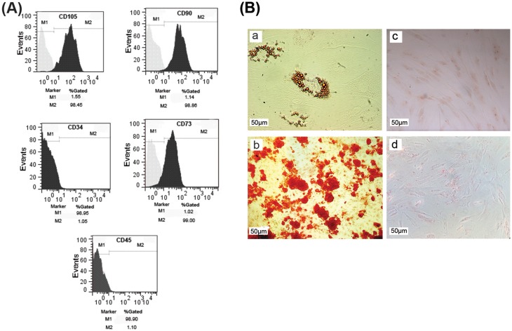
Methods: LPC (1%, 2 µL) was injected into the rat optic chiasm and animals underwent daily intraperitoneal (i.p.) injections of ADSCs-CM and oral gavage of aspirin at a dose of 25 mg/kg for 14 days post LPC injection. The conductivity of visual signals was assessed using visual evoked potential recordings (VEPs) before LPC injection and on days 7 and 14 post lesion. Immunostaining against PDGFRα as oligodendrocyte precursor cells marker, MOG as mature myelin marker, and GFAP as astrocyte marker was performed on brain sections at day 14 post LPC injection. FluoroMyelin staining was also used to measure the extent of demyelination areas.

Results: Our results showed that administration of ADSCs-CM and aspirin significantly reduced the latency of VEP waves in LPC receiving animals. In addition, demyelination levels and GFAP expressing cells were attenuated while the number of oligodendrocyte precursor cells significantly increased in rats treated with ADSCs-CM and aspirin.

Conclusion: Overall, our results suggest that co-administration of ADSCs-CM and aspirin improves the functional recovery of optic pathway through amelioration of astrocyte activation and attenuation of demyelination level.

Keywords: aspirin; conditioned medium; demyelination; lysolecithin; mesenchymal stem cells; optic chiasm.


Conflict of interest statement

The authors declare no conflict of interest related to this study.

Figures

Figure 1
Figure 1
Characterization of cultured ADSCs. (A) Flow cytometry analysis showed that cultured cells expressed the specific markers of ADSCs such as CD105, CD90, and CD73, while they were negative for CD34 and CD45 as hematopoietic stem cells markers. (B) Differentiation capacity of ADSCs into adipocytes (a) and osteoblasts (b); (c, d) undifferentiated cells as negative control. Scale bar: 50 µm.
Figure 2
Figure 2
Effect of ADSCs-CM and aspirin on conductivity of visual signals. (A) Sample traces of VEP waves in experimental groups. Scale bar: voltage: 10 µV, time: 50 ms. (B) Quantification of VEP data indicated that co-administration of ADSCs-CM and aspirin improved the functional recovery of optic pathway. Two-way ANOVA followed by Bonferroni post-test were used for statistical analysis. **P<0.01 and ***P<0.001 compared to the saline + saline; P<0.001 compared to the LPC + vehicle; &&P<0.01 and &&&P<0.001 compared to the LPC + aspirin and $P<0.05 compared to the LPC + ADSCs-CM. n=6. Abbreviation: Dpi, days post injection.
Figure 3
Figure 3
Effect of ADSCs-CM and aspirin on demyelination levels of the optic chiasm. (A) FluoroMyelin staining of optic chiasm’s sections on day 14 post LPC injection. DAPI: nucleus stain. Scale bar: 100 µm. (B) ADSCs-CM, aspirin or combination of ADSCs-CM and aspirin decreased the extent of demyelination areas. One-way ANOVA followed by Tukey post-test were used for statistical analysis. **P<0.01 and ***P<0.001 compared to LPC + vehicle. n=3.
Figure 4
Figure 4
Effect of ADSCs-CM and aspirin on extent of demyelination areas. (A) Immunostaining against MOG as mature myelin marker on day 14 post insult. DAPI: nuclei stain, scale bar: 100 µm. (B) Quantification of MOG staining showed that administration of ADSCs-CM, aspirin or ADSCs-CM + aspirin alleviated the level of demyelination. One-way ANOVA followed by Tukey post-test were used for statistical analysis. ***P<0.001 compared to the LPC + vehicle; #P<0.05 compared to the LPC + ADSCs-CM and &&P<0.01 compared to the LPC + aspirin. n=3.
Figure 5
Figure 5
Effect of ADSCs-CM and aspirin on the number of oligodendrocyte precursor cells. (A) Immunostaining against PDGFRα as oligodendrocyte precursor cells marker on brain sections. DAPI: nuclei stain. Scale bar: 100 µm. (B) Quantification of immunostaining data showed that co-administration of ADSCs-CM and aspirin increased the number of PDGFRα expressing cells following LPC injection. One-way ANOVA followed by Tukey post-test were used for statistical analysis. ***P<0.001 compared to the saline + saline; P<0.001 compared to the LPC + vehicle; &&P<0.01 compared to the LPC + ADSCs-CM and $$P<0.01 compared to the LPC + aspirin. n=3.
Figure 6
Figure 6
Effect of ADSCs-CM and aspirin on astrocyte activation of optic chiasm in LPC-induced demyelination model. (A) Immunostaining against GFAP as astrocyte marker on day 14 post LPC injection. DAPI: nuclei stain. Scale bar: 100 µm. (B) ADSCs-CM, aspirin or ADSCs-CM + aspirin ameliorated the number of GFAP positive cells in the demyelination context. One-way ANOVA followed by Tukey post-test were used for statistical analysis. *P<0.05 and ***P<0.001 compared to the saline + saline and P<0.001 compared to the LPC + vehicle. n=3.

References

    1. Ghasemi‐Kasman M, Zare L, Baharvand H, Javan M. In vivo conversion of astrocytes to myelinating cells by miR-302/367 and valproate to enhance myelin repair. J Tissue Eng Regen Med. 2018;12(1):e462–e472. doi:10.1002/term.2276 - DOI - PubMed
    1. Yousefi F, Ebtekar M, Soudi S, Soleimani M, Hashemi SM. In vivo immunomodulatory effects of adipose-derived mesenchymal stem cells conditioned medium in experimental autoimmune encephalomyelitis. Immunol Lett. 2016;172:94–105. doi:10.1016/j.imlet.2016.02.016 - DOI - PubMed
    1. Mondal S, Jana M, Dasarathi S, Roy A, Pahan K. Aspirin ameliorates experimental autoimmune encephalomyelitis through interleukin-11–mediated protection of regulatory T cells. Sci Signal. 2018;11(558):eaar8278. doi:10.1126/scisignal.aar8278 - DOI - PMC - PubMed
    1. Mulakayala N, Rao P, Iqbal J, Bandichhor R, Oruganti S. Synthesis of novel therapeutic agents for the treatment of multiple sclerosis: a brief overview. Eur J Med Chem. 2013;60:170–186. doi:10.1016/j.ejmech.2012.10.055 - DOI - PubMed
    1. Jadasz JJ, Kremer D, Göttle P, et al. Mesenchymal stem cell conditioning promotes rat oligodendroglial cell maturation. PLoS One. 2013;8(8):e71814. doi:10.1371/journal.pone.0071814 - DOI - PMC - PubMed