. 2019 Sep 12:12:691-698.
doi: 10.2147/CCID.S223317. eCollection 2019.

Erosive pustular dermatosis of the scalp: challenges and solutions

Affiliations

Erosive pustular dermatosis of the scalp: challenges and solutions

Michela Starace et al. Clin Cosmet Investig Dermatol. .

Abstract

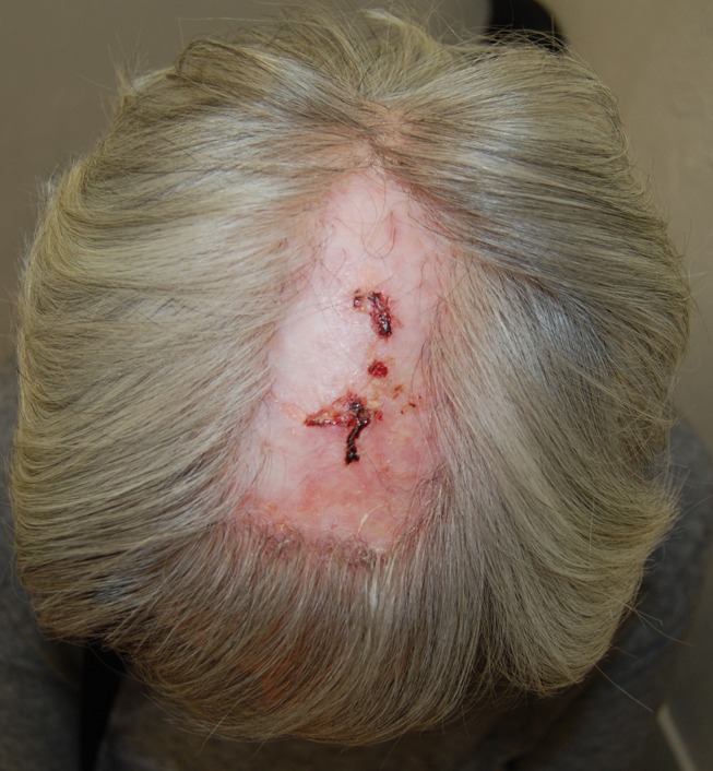
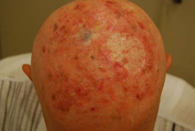
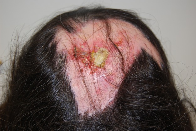
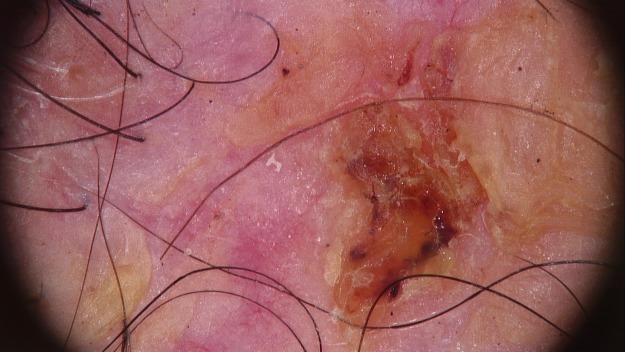
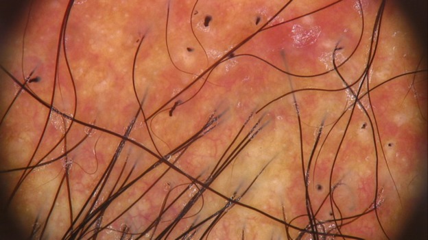
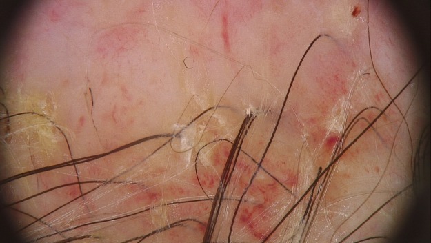
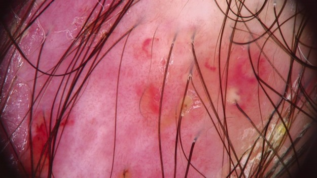
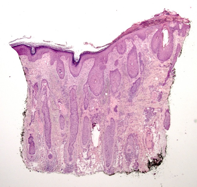
Erosive pustular dermatosis of the scalp is a rare chronic inflammatory disorder defined. It usually affects elderly people and is characterized by extensive pustular lesions, erosions, and crusts located on the scalp. The pathogenesis of this disease is not completely understood, but a known predisposing factor is skin trauma. Autoimmune disorders including rheumatoid arthritis, autoimmune hepatitis, Hashimoto thyroiditis, and Takayasu aortitis are associated diseases reported. The clinical examination reveals erythema, erosions, crusts, follicular pustules, and in advanced stages, scarring alopecia. A scalp biopsy is recommended but not specific, founding epidermal atrophy, focal erosions, and a mixed inflammatory infiltrate consisting of neutrophils, lymphocytes, and plasma cells. Bacterial cultures, fungal and viral stains are not necessary and are usually negative. . Topical high-potency corticosteroids, retinoids, calcipotriol, dapsone, and topical tacrolimus are reported treatments, while photodynamic therapy has been effective in some patients, but has induced the disease in others. All the findings are suggestive but not specific, so it is an excluding diagnosis. The combination of predisposing factors is very important for a correct diagnosis, such as elderly age, sun-damaged skin, presence of androgenetic alopecia, together with clinical manifestations, non-specific histology and laboratory investigations negative for other disease. In our opinion, this scalp disease is a challenge for the dermatologist. We review all the literature to better define the possible solutions in case of suspected erosive pustular dermatosis of the scalp.

Keywords: cicatricial alopecia; scalp erosions; scalp trauma; scarring alopecia; trichoscopy.


Conflict of interest statement

The authors report no conflicts of interest in this work.

Figures

Figure 1
Figure 1
Typical clinical aspect of EPDS with superficial erosions and crusted lesions on atrophic skin. Abbreviation: EPDS, erosive pustular dermatosis of the scalp.
Figure 2
Figure 2
Actinic damage and epidermal atrophy can predispose to EPDS. Abbreviation: EPDS, erosive pustular dermatosis of the scalp.
Figure 3
Figure 3
A severe case of EPDS large, erosive, and crusted patches with overlying and surrounding pustules and serum exudate involving the bald areas of the scalp at the vertex. Abbreviation: EPDS, erosive pustular dermatosis of the scalp.
Figure 4
Figure 4
Trichoscopy picture showing absence of follicular openings, hemorrhagic crusts, and hair shaft tortuosity.
Figure 5
Figure 5
Active phase of the disease with red and milky areas, absence of follicular opening, and several hair shafts broken.
Figure 6
Figure 6
Trichoscopy of chronic EPDS where there is evident marked atrophic skin, lack of follicular ostia, and follicular yellow thick exudate. Abbreviation: EPDS, erosive pustular dermatosis of the scalp.
Figure 7
Figure 7
The hypergranulation variant of EPDS showing different types of vessels at trichoscopy. Abbreviation: EPDS, erosive pustular dermatosis of the scalp.
Figure 8
Figure 8
Early stage of erosive pustular dermatosis of the scalp: pathology shows laminated orthokeratosis and psoriasiform hyperplasia at the epidermis; slight mixed inflammatory infiltrate mainly consisting of neutrophils, lymphocytes, and plasmocytes, and mild fibrosis at the papillary dermis; hair follicular density is normal, with terminal, miniaturized anagen follicles and an increased number of catagen follicles.

Similar articles

Cited by

References

    1. Patton D, Lynch PJ, Fung MA, Fazel N. Chronic atrophic erosive dermatosis of the scalp and extremities: a recharacterisation of erosive pustular dermatosis. J Am Acad Dermatol. 2007;57:421. doi:10.1016/j.jaad.2007.04.026 - DOI - PubMed
    1. Starace M, Loi C, Bruni F, et al. Erosive pustular dermatosis of the scalp: clinical, trichoscopic, and histopathologic features of 20 cases. J Am Acad Dermatol. 2017;76(6):1109–1114.e2. doi:10.1016/j.jaad.2016.12.016 - DOI - PubMed
    1. Fertig R, Maddy A, Cadore de Faris D, Simao Correa Fiho S, Rigatti M, Tosti A. Erosive pustular dermatosis of the scalp after aplasia cutis congenita in a 9-year-old patient: a 5-year follow-up. Pediatr Dermatol. 2017;34:695–696. doi:10.1111/pde.13252 - DOI - PubMed
    1. LaCour M, Allen T, Wilkerson M, Nguyen AV. A case of erosive pustular dermatosis in a pediatric patient. JAAD Case Rep. 2019;5:118–120. doi:10.1016/j.jdcr.2018.11.001 - DOI - PMC - PubMed
    1. Burton JL. Case for diagnosis. Pustular dermatosis of the scalp. Br J Dermatol. 1977;97 Suppl:15-67-9. - PubMed

LinkOut - more resources