. 2019 Sep 18:11:8507-8516.
doi: 10.2147/CMAR.S212380. eCollection 2019.

Clinicopathological features and outcomes of esophageal lesions containing a basal layer type squamous cell carcinoma component

Affiliations

Clinicopathological features and outcomes of esophageal lesions containing a basal layer type squamous cell carcinoma component

Xiaoduan Zhuang et al. Cancer Manag Res. .

Abstract

Purpose: Basal layer type squamous cell carcinoma (BLSCC) is a unique type of squamous cell carcinoma (SCC), characterized by high-grade dysplastic cells occupying the lower half of the epithelium. So far, such special lesions do not seem to attract much attention. The aim of this study was to investigate the clinicopathological features and prognosis of esophageal squamous carcinoma lesions with a BLSCC component.

Materials and methods: Between January 2011 and January 2018, 96 patients with esophageal squamous cell carcinoma underwent endoscopic submucosal resection in our hospital were retrospectively analyzed. Patients were divided into BLSCC or typical SCC groups according to the presence or absence of a BLSCC component. The endoscopic findings were compared between the two groups. Furthermore, patients were followed up until October 2018 to compare recurrence rates.

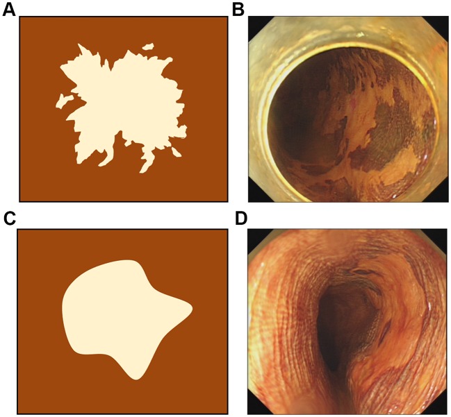
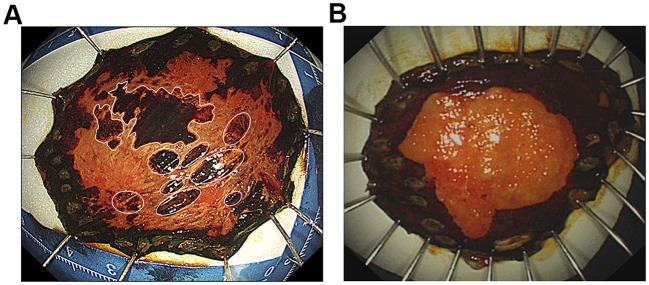
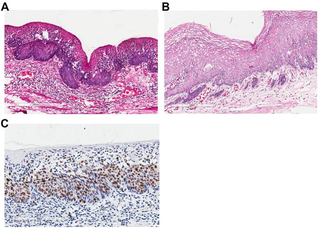
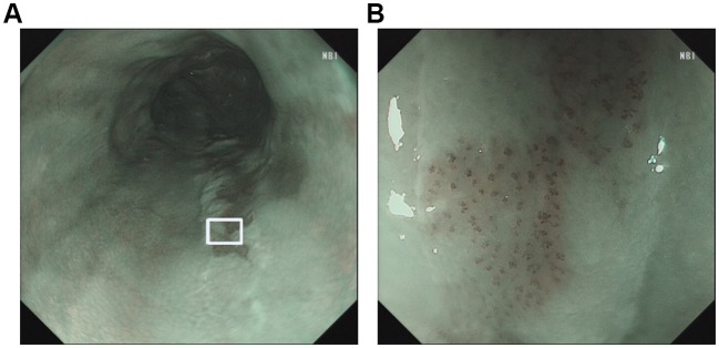
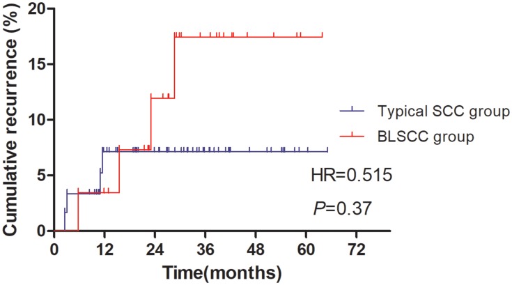
Results: BLSCC components were detected in 32 (33.3%, 32/96) lesions. Among them, 13 (40.62%, 13/32) were BLSCC predominant. The intraepithelial papillary capillary loops of 7 pure BLSCC showed type B1 under narrow-band imaging. Single-factor and multivariate analyses indicated that five or more independently scattered, deep-stained spots in iodine-unstained areas were significantly predictive of the presence of BLSCC components (OR=4.837, P=0.015). All patients of typical SCC group survived, but one of BLSCC group died for distant metastases during the follow-up period. The 1-year cumulative recurrence rate (CRR) of BLSCC group were 3.4%, lower than that of typical SCC group (7.1%). Although no significant difference of CRR was seen between the two groups (P>0.05), the 2-year CRR of BLSCC group increased to 11.9%, being higher than that of typical SCC group (7.1%).

Conclusion: The presence of multiple, scattered stained spots in iodine-unstained areas was predictive of BLSCC components. Such lesion should be treated actively and subject to a more rigorous follow-up protocol due to a higher likelihood of late recurrence.

Keywords: basal layer type squamous cell carcinoma; clinicopathology; esophagus; recurrence.


Conflict of interest statement

The authors report no conflicts of interest in this work.

Figures

Figure 1
Figure 1
Images of esophageal squamous epithelial neoplastic lesions after iodine staining. (A) A schematic drawing of the worm-eaten boundary of an iodine-unstained area (irregular type). (B) A lesion with an irregular type of boundary of an iodine-unstained area. (C) A schematic drawing of the smooth boundary of an iodine-unstained area (regular type). (D) A lesion with a regular type and a well-demarcated boundary of an iodine-unstained area.
Figure 2
Figure 2
Images of endoscopic submucosal resection specimens stained by Lugol dye solution. (A) A lesion with more than five stained spots (marked by white circles), which were independently scattered in the iodine-unstained area, without connection to the surrounding iodine-stained area, was defined as positive for multiple stained spots in an iodine-free area. (B) A lesion with less than five scattered, deep-stained spots in the iodine-unstained area; negative for multiple stains in the iodine-free area.
Figure 3
Figure 3
The invasive growth patterns and Ki-67 immunohistological findings of BLSCC components. (A) The BLSCC component presented with an infiltrative growth pattern, INFa, toward the LPM. (B) The BLSCC component exhibited INFb with minimal tumor foci in the LPM. (C) Ki-67 positive cells were mainly detected in the lower half of the epithelial layer of BLSCC components. Abbreviations: LPM, lamina propria mucosae; INF, infiltration growth pattern.
Figure 4
Figure 4
The NBI images of BLSCC. (A) Endoscopic image under NBI showed the lesion with brownish color change. (B) A magnified view of the white box area in Figure A. The IPCLs exhibited the changes of dilatation, meandering, irregular caliber, and form variation, belonging to JES type B1. Abbreviations: NBI, narrow-band imaging; IPCL, intraepithelial papillary capillary loop; JES, Japan Esophageal Society.
Figure 5
Figure 5
Comparison of cumulative recurrence rates between BLSCC group and typical SCC group.

Similar articles

Cited by

References

    1. Japan Esophageal Society. Japanese classification of esophageal cancer, 11th edition: part II and III. Esophagus-Tokyo 2017;14(1):37–65. doi:10.1007/s10388-016-0556-2. - DOI - PMC - PubMed
    1. Japanese Society of Esophageal Diseases. [Guidelines for the Clinical and Pathologic Studies on Carcinoma of the Esophagus] 9th edn. Tokyo: Kanehara, 1999. (In Japanese.).
    1. Schlemper RJ, Kato Y, Stolte M. Diagnostic criteria for gastrointestinal carcinomas in Japan and Western countries: proposal for a new classification system of gastrointestinal epithelial neoplasia. J Gastroenterol Hepatol. 2010;15(s3):G49–G57. doi:10.1046/j.1440-1746.2000.02266.x - DOI - PubMed
    1. Schlemper RJ, Kato Y, Stolte M. Review of histological classifications of gastrointestinal epithelial neoplasia: differences in diagnosis of early carcinomas between Japanese and Western pathologists. J Gastroenterol. 2001;36(7):445–456. doi:10.1007/s005350170067 - DOI - PubMed
    1. Shimizu M, Nagata K, Yamaguchi H, Kita H. Squamous intraepithelial neoplasia of the esophagus: past, present, and future. J Gastroenterol. 2009;44(2):103–112. doi:10.1007/s00535-008-2298-y - DOI - PubMed