Functional-Optical Coherence Tomography: A Non-invasive Approach to Assess the Sympathetic Nervous System and Intrinsic Vascular Regulation
- PMID: 31572206
- PMCID: PMC6751282
- DOI: 10.3389/fphys.2019.01146
Functional-Optical Coherence Tomography: A Non-invasive Approach to Assess the Sympathetic Nervous System and Intrinsic Vascular Regulation
Abstract
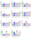
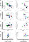
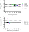
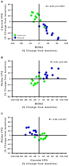
Sympathetic nervous system dysregulation and vascular impairment in neuronal tissue beds are hallmarks of prominent cardiorespiratory diseases. However, an accurate and convenient method of assessing SNA and local vascular regulation is lacking, hindering routine clinical and research assessments. To address this, we investigated whether spectral domain optical coherence tomography (OCT), that allows investigation of retina and choroid vascular responsiveness, reflects sympathetic activity in order to develop a quick, easy and non-invasive sympathetic index. Here, we compare choroid and retina vascular perfusion density (VPD) acquired with OCT and heart rate variability (HRV) to microneurography. We recruited 6 healthy males (26 ± 3 years) and 5 healthy females (23 ± 1 year) and instrumented them for respiratory parameters, ECG, blood pressure and muscle sympathetic nerve microneurography. Choroid VPD decreases with the cold pressor test, inhaled hypoxia and breath-hold, and increases with hyperoxia and hyperpnea suggesting that sympathetic activity dominates choroid responses. In contrast, retina VPD was unaffected by the cold pressor test, increased with hypoxia and breath hold and decreases with hyperoxia and hyperpnea, suggesting metabolic vascular regulation dominates the retina. With regards to integrated muscle sympathetic nerve activity, HRV had low predictive power whereas choroid VPD was strongly (inversely) correlated with integrated muscle sympathetic nerve activity (R = -0.76; p < 0.0001). These data suggest that Functional-OCT may provide a novel approach to assess sympathetic activity and intrinsic vascular responsiveness (i.e., autoregulation). Given that sympathetic nervous system activity is the main determinant of autonomic function, sympathetic excitation is associated with severe cardiovascular/cardiorespiratory diseases and autoregulation is critical for brain health, we suggest that the use of our new Functional-OCT technique will be of broad interest to clinicians and researchers.
Keywords: autoregulation; choroid; microvascular; sympathetic nervous system; vascular.
Copyright © 2019 Jendzjowsky, Steinback, Herman, Tsai, Costello and Wilson.
Figures


Similar articles
-
Carbohydrate ingestion induces differential autonomic dysregulation in normal-tension glaucoma and primary open angle glaucoma.PLoS One. 2018 Jun 7;13(6):e0198432. doi: 10.1371/journal.pone.0198432. eCollection 2018. PLoS One. 2018. PMID: 29879162 Free PMC article.
-
Autonomic nerve and cardiovascular responses to changing blood oxygen and carbon dioxide levels in the rat.J Auton Nerv Syst. 1989 Oct;28(1):61-74. doi: 10.1016/0165-1838(89)90008-8. J Auton Nerv Syst. 1989. PMID: 2511237
-
Anodal transcranial direct current stimulation (tDCS) over the motor cortex increases sympathetic nerve activity.Brain Stimul. 2014 Jan-Feb;7(1):97-104. doi: 10.1016/j.brs.2013.08.005. Epub 2013 Sep 18. Brain Stimul. 2014. PMID: 24080439 Clinical Trial.
-
[Heart rate variability. Applications in psychiatry].Encephale. 2009 Oct;35(5):423-8. doi: 10.1016/j.encep.2008.06.016. Epub 2008 Dec 18. Encephale. 2009. PMID: 19853714 Review. French.
-
Noradrenaline spillover and microneurography measurements in patients with primary hypertension.J Hypertens Suppl. 1998 Aug;16(3):S35-8. J Hypertens Suppl. 1998. PMID: 9747908 Review.
Cited by
-
The short-term effects of blood donation on the ocular parameters including blood flow of the retina and choroid in healthy people using OCT- angiography.BMC Ophthalmol. 2023 Jun 12;23(1):259. doi: 10.1186/s12886-023-03002-3. BMC Ophthalmol. 2023. PMID: 37303035 Free PMC article.
-
Choroidal Thickness Increases During Parasympathetic Dominance After Immersion of the Foot in Warm Water.Cureus. 2024 Jan 29;16(1):e53194. doi: 10.7759/cureus.53194. eCollection 2024 Jan. Cureus. 2024. PMID: 38425624 Free PMC article.
-
An Integrative Literature Review of Heart Rate Variability Measures to Determine Autonomic Nervous System Responsiveness Using Pharmacological Manipulation.J Cardiovasc Nurs. 2024 Jan-Feb 01;39(1):58-78. doi: 10.1097/JCN.0000000000001001. Epub 2023 May 27. J Cardiovasc Nurs. 2024. PMID: 37249528 Free PMC article. Review.
-
The effect of hyperoxia on muscle sympathetic nerve activity: a systematic review and meta-analysis.Clin Auton Res. 2024 Apr;34(2):233-252. doi: 10.1007/s10286-024-01033-4. Epub 2024 May 6. Clin Auton Res. 2024. PMID: 38709357
-
Choroidal Morphology and Systemic Circulation Changes During the Menstrual Cycle in Healthy Japanese Women.Cureus. 2023 Nov 1;15(11):e48124. doi: 10.7759/cureus.48124. eCollection 2023 Nov. Cureus. 2023. PMID: 38046755 Free PMC article.
References
-
- Baumert M., Lambert G. W., Dawood T., Lambert E. A., Esler M. D., McGrane M., et al. (2009). Short-term heart rate variability and cardiac norepinephrine spillover in patients with depression and panic disorder. Am. J. Physiol. Heart Circ. Physiol. 297 H674–H679. 10.1152/ajpheart.00236.2009 - DOI - PubMed
LinkOut - more resources
Full Text Sources
Research Materials

