. 2019 Sep 12:10:971.
doi: 10.3389/fneur.2019.00971. eCollection 2019.

FK506 Attenuated Pilocarpine-Induced Epilepsy by Reducing Inflammation in Rats

Affiliations

FK506 Attenuated Pilocarpine-Induced Epilepsy by Reducing Inflammation in Rats

Aihua Wang et al. Front Neurol. .

Abstract

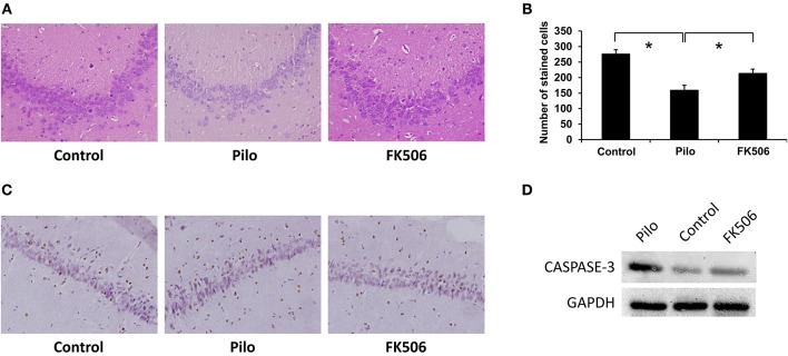
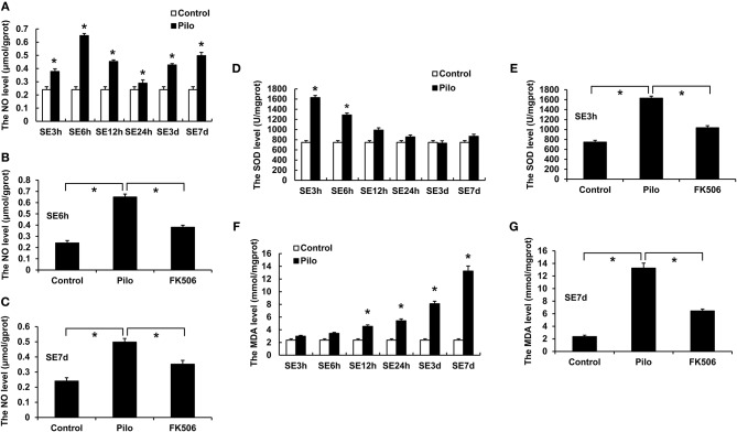
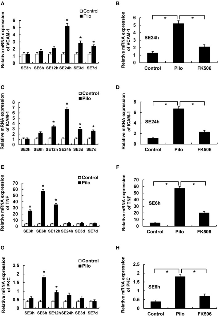
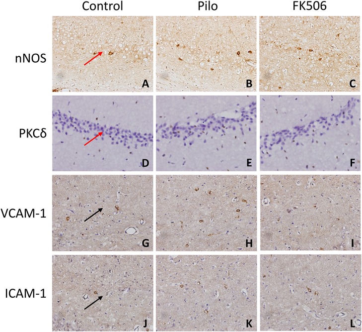
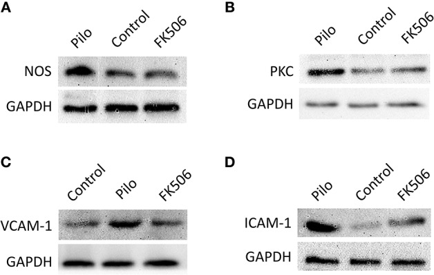
Background: The status epilepticus (SE) is accompanied by a local inflammatory response and many oxygen free radicals. FK506 is an effective immunosuppressive agent with neuroprotective and neurotrophic effects, however, whether it can inhibit the inflammatory response and attenuate epilepsy remains unclear. Objective: This study aims to clarify the effect of FK506 on inflammatory response in rats with epilepsy. Methods: A total of 180 rats were randomly and equally divided into the control group, epilepsy group, and FK506 group. The rat SE model in the epilepsy group and FK506 group was induced by lithium chloride combined with pilocarpine. In the FK506 group, FK506 was given before the injection of pilocarpine. The control group was given the same volume of saline. Then the effect of FK506 on epilepsy in rats and the changes of inflammatory factors and free radicals in hippocampus were examined using hematoxylin and eosin (HE) staining, immunohistochemistry, quantitative real-time polymerase chain reaction (qRT-PCR), and western blotting. Results: FK506 ameliorated the course of pilocarpine-induced epilepsy and the neuronal loss in the rat hippocampus after SE. FK506 reduced the increased content of nitric oxide (NO), superoxide dismutase (SOD), and malondialdehyde (MDA) in the hippocampus after SE. Besides, FK506 also significantly reduced the levels of factors involved in inflammatory response such as vascular cell adhesion molecule-1 (VCAM-1), intercellular adhesion molecule-1 (ICAM-1), tumor necrosis factor-α (TNF-α), and Protein Kinase C δ (PKCδ) that rise after epilepsy. Conclusion: FK506 ameliorated the course of pilocarpine-induced epilepsy, significantly reduced free radical content, and inhibited the expression of inflammatory factors, which provided a theoretical basis for the application of FK506 in the treatment of epilepsy.

Keywords: FK506; inflammatory response; neuroprotective; radical content; status epilepsy.


Figures

Figure 1
Figure 1
Changes of neuronal loss in the hippocampus of rats in different treatment groups. (A) Morphological changes (HE staining) of the hippocampus in different treatment groups after 7 days of modeling. Magnification: 400 X. (B) Unbiased quantification of HE stained pyramidal CA3 cells. *P < 0.05. (C) Caspase-3 expression in the control group, epilepsy group, and FK506 group after 7 days of modeling detected by immunohistochemistry. Magnification: 400 X. (D) Protein expression of Caspase-3 in the control group, epilepsy group, and FK506 group after 7 days of modeling detected by western blotting.
Figure 2
Figure 2
Changes of NO, MDA, and SOD in the hippocampus of rats in different treatment groups. (A) Changes of NO at different time points in the control group and the epilepsy group. (B) Changes of NO in the control group, epilepsy group, and FK506 group after 6 h of modeling. (C) Changes of NO in the control group, epilepsy group, and FK506 group after 7 days of modeling. (D) Changes of SOD at different time points in the control group and the epilepsy group. (E) Changes of SOD in the control group, epilepsy group, and FK506 group after 3 h of modeling. (F) Changes of MDA at different time points in the control group and the epilepsy group. (G) Changes of MDA in the control group, epilepsy group, and FK506 group after 7 days of modeling. *P < 0.05.
Figure 3
Figure 3
The RNA levels of VCAM-1, ICAM-1, PKCδ, and TNF in the hippocampus of different treatment groups. (A) VCAM-1 changes at different time points in the control group and the epilepsy group. (B) VCAM-1 changes in the control group, epilepsy group, and FK506 group after 24 h of modeling. (C) ICAM-1 changes at different time points in the control group and the epilepsy group. (D) ICAM-1 changes in the control group, epilepsy group, and FK506 group after 24 h of modeling. (E) TNF-α changes at different time points in the control group and the epilepsy group. (F) TNF-α changes in the control group, epilepsy group, and FK506 group after 6 h of modeling. (G) PKCδ changes at different time points in the control group and the epilepsy group. (H) PKCδ changes in the control group, epilepsy group, and FK506 group after 6 h of modeling. *P < 0.05.
Figure 4
Figure 4
The in situ expression of VCAM-1, ICAM-1, PKCδ, NOS, and caspase-3 in the hippocampus of different treatment groups. (A–C) NOS expression in the control group, epilepsy group, and FK506 group after 6 h of modeling. (D–F) PKCδ expression in the control group, epilepsy group, and FK506 group after 6 h of modeling. (G–I) VCAM-1 expression in the control group, epilepsy group, and FK506 group after 24 h of modeling. (J–L) ICAM-1 expression in the control group, epilepsy group, and FK506 group after 24 h of modeling. Magnification: 400 X. Red arrow, CA3 region. Black arrow, vascular endothelium.
Figure 5
Figure 5
The protein expression of VCAM-1, ICAM-1, PKCδ, and TNF in the hippocampus of different treatment groups. (A) Protein expression of NOS in the control group, epilepsy group, and FK506 group after 6 h of modeling. (B) Protein expression of PKCδ in the control group, epilepsy group, and FK506 group after 6 h of modeling. (C) Protein expression of VCAM-1 in the control group, epilepsy group, and FK506 group after 24 h of modeling. (D) Protein expression of ICAM-1 in the control group, epilepsy group, and FK506 group after 24 h of modeling. The internal reference was glyceraldehyde-3-phosphate dehydrogenase (GAPDH).

Similar articles

Cited by

References

    1. Devinsky O, Vezzani A, O'Brien TJ, Jette N, Scheffer IE, de Curtis M, et al. Epilepsy. Nat Rev Dis Primers. (2018) 4:18024 10.1038/nrdp.2018.24 - DOI - PubMed
    1. Motamedi G, Meador K. Epilepsy and cognition. Epilepsy Behav. (2003) 4(Suppl 2):S25–38. 10.1016/j.yebeh.2003.07.004 - DOI - PubMed
    1. Rodriguez-Alvarez N, Jimenez-Mateos EM, Dunleavy M, Waddington JL, Boylan GB, Henshall DC. Effects of hypoxia-induced neonatal seizures on acute hippocampal injury and later-life seizure susceptibility and anxiety-related behavior in mice. Neurobiol Dis. (2015) 83:100–14. 10.1016/j.nbd.2015.08.023 - DOI - PubMed
    1. Marawar R, Basha M, Mahulikar A, Desai A, Suchdev K, Shah A. Updates in refractory status epilepticus. Crit Care Res Pract. (2018) 2018:9768949. 10.1155/2018/9768949 - DOI - PMC - PubMed
    1. Trinka E, Cock H, Hesdorffer D, Rossetti AO, Scheffer IE, Shinnar S, et al. . A definition and classification of status epilepticus–report of the ILAE Task force on classification of status epilepticus. Epilepsia. (2015) 56:1515–23. 10.1111/epi.13121 - DOI - PubMed