Review
. 2019 Sep 11:10:2138.
doi: 10.3389/fimmu.2019.02138. eCollection 2019.

Factors Affecting Early Antibody Secreting Cell Maturation Into Long-Lived Plasma Cells

Affiliations
Review

Factors Affecting Early Antibody Secreting Cell Maturation Into Long-Lived Plasma Cells

Doan C Nguyen et al. Front Immunol. .

Abstract

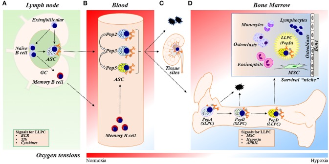
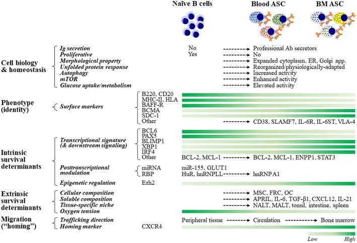
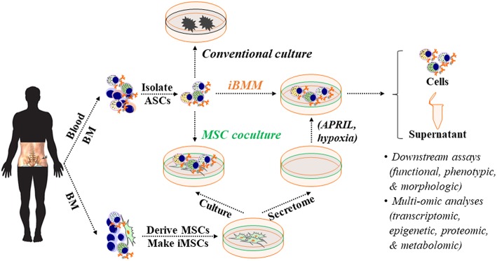
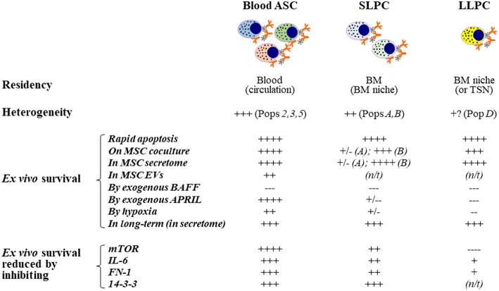
Antibody secreting cells (ASCs) are terminally differentiated cells of the humoral immune response and must adapt morphologically, transcriptionally, and metabolically to maintain high-rates of antibody (Ab) secretion. ASCs differentiate from activated B cells in lymph nodes and transiently circulate in the blood. Most of the circulating ASCs undergo apoptosis, but a small fraction of early ASCs migrate to the bone marrow (BM) and eventually mature into long-lived plasma cells (LLPCs). LLPC survival is controlled both intrinsically and extrinsically. Their differentiation and maintenance programs are governed by many intrinsic mechanisms involving anti-apoptosis, autophagy, and metabolism. The extrinsic factors involved in LLPC generation include BM stromal cells, cytokines, and chemokines, such as APRIL, IL-6, and CXCL12. In humans, the BM CD19-CD38hiCD138+ ASC subset is the main repository of LLPCs, and our recent development of an in vitro BM mimic provides essential tools to study environmental cues that support LLPC survival and the critical molecular mechanisms of maturation from early minted blood ASCs to LLPCs. In this review, we summarize the evidence of LLPC generation and maintenance and provide novel paradigms of LLPC maturation.

Keywords: B cell; antibody-secreting cell; differentiation; immunoglobulin; long-lived plasma cell; maintenance; maturation.


Figures

Figure 1
Figure 1
ASC differentiation, maturation, and LLPC generation and maintenance. (A) In response to vaccination or infection, naïve B cells proliferate and differentiate into ASCs within germinal center (GC) reactions in secondary lymphoid organs (SLOs) such as lymph nodes (LNs) or outside of GCs in extrafollicular responses. ASCs then egress out of SLOs and circulate in the blood transiently. Naïve B cells can also differentiate into memory B cells, circulate systemically, and further differentiate into ASCs upon activation. The two-step model of LLPC generation consists of the GC reaction and the BM maturation process [see also (D)]. In the GC reaction, B cells may receive signals such as BCR stimulation, Tfh (or other Th cell) interactions, and cytokines (such as IL-21 and IFNγ) in the GC that are important during ASC differentiation to ultimately become a LLPC. (B) Human blood ASCs are heterogeneous and can be classified into distinct populations (Pops), including Pop2, Pop3, and Pop5, based on their surface protein expression (see also Table 1). (C) Most blood ASCs apoptose. However, a fraction migrates to inflammatory or other tissue “niches” (e.g., mucosa and bone marrow) and become tissue-specific ASCs. (D) A small number of blood ASCs emigrate to the BM, a physiologically hypoxic environment. Human BM ASCs can be identified based on surface protein expression as PopA (SLPCs), PopB (SLPCs), and PopD (LLPCs), respectively, in the BM (Table 1). LLPCs acquire longevity and are maintained within the dedicated BM survival “niche.” ASC survival factors include BM MSCs (and/or their secreted factors), cytokines (i.e., APRIL), and hypoxic conditions. BM, bone marrow; GC, germinal center; ASC, antibody-secreting cells; SLPC, short-lived plasma cells; LLPC, long-lived plasma cells; BCR, B-cell receptor; Tfh, T follicular helper cells; MSC, mesenchymal stromal/stem cells.
Figure 2
Figure 2
ASC cell biology, fate determination, and resultant phenotypes. ASCs adapt their cellular biology, machinery, and morphology to maintain homeostasis while producing copious amounts of antibodies. For example, ASCs must upregulate the unfolded protein response (UPR) to inhibit apoptotic pathways that would be engaged due to increased Ig synthesis. Other pathways that act in concert with the UPR to support ASC homeostasis are autophagy, mTOR, and glucose metabolism. Although no phenotype-based “universal identity” of blood and bone marrow ASCs presently exists, ASCs are typically identified by increased expression of CD38, CD138, and BCMA, and decreased expression of B220, MHCII, and BCR. The commitment to the ASC fate of naïve B cells is regulated transcriptionally, post-transcriptionally, and epigenetically. Maintenance of B cell programs is mainly controlled by Bcl6 and Pax5, and promotion of B cell differentiation and ASC commitment are essentially governed by Blimp1 (encoded by Prdm1), Xbp1, and Irf4. The role of post-transcriptional (through certain miRNAs and RBPs) and epigenetic (such as via Ezh2) regulation is increasingly being recognized. The lifespan of ASCs ranges from a few days to several decades and is thought to be chiefly determined extrinsically, which is largely dependent on their residency. Accordingly, LLPCs reside in a specialized, dedicated BM compartment (“survival niche”) that is physiologically hypoxic and is formed and maintained by both cellular (such as MSCs, FRCs, and OCs) and soluble (such as APRIL, IL-6, TGF-β1, and CXCL12) components. Other tissue-specific niches capable of maintaining ASCs have also been described. ASC migration (or “homing”) from peripheral tissues or SLOs toward survival niches is primarily directed by binding of CXCR4 to its ligand, MSC-secreted chemokine CXCL12 (SDF1α). Certain miRNAs and RBPs include miR-155, which is involved in cytokine production, and HuR and hnRNPLL, which modulate IgH mRNA turnover and translation. ASC, antibody-secreting cells; LLPC, long-lived plasma cells; ER, endoplasmic reticulum; SLO, secondary lymphoid organs; MSC, mesenchymal stromal cells; FRC, fibroblastic reticular cells; OC, osteoclasts; NALT, nasal-associated lymphoid tissues; MALT, mucosa-associated lymphoid tissues.
Figure 3
Figure 3
An in vitro BM microniche mimic (iBMM) for studying human ASCs ex vivo. Primary MSCs are derived from human autologous or allogeneic BM samples, expanded, and irradiated. Populations or single blood or BM ASCs are isolated and co-cultured with BM-derived iMSCs or in the cell-free secretome that is harvested from BM-derived MSCs. Typically, blood or BM ASCs quickly die off ex vivo or in conventional culture but survive for weeks under these conditions. The cellular materials and supernatants obtained from the ex vivo ASC cultures can be collected for downstream assays and multi-omic analyses for studying human ASC biology. BM, bone marrow; ASC, antibody-secreting cells; MSC, mesenchymal stromal/stem cells; iMSC, irradiated MSC; APRIL, a proliferation-inducing ligand.
Figure 4
Figure 4
Extrinsic cues for human ex vivo ASC maintenance: Findings from using the in vitro human BM mimic. Blood ASCs circulate transiently while short- and long-lived PCs reside in tissue-specific niches like the BM. Both circulating ASC and local PC populations are heterogeneous in humans and can be classified into distinct subsets based on their expression of CD19, CD38, and CD138. These populations are named Pop2, Pop3, or Pop5 in blood and PopA, PopB, or PopD in the BM. Typically, each of these ASC populations undergo rapid apoptosis ex vivo; however, they can survive for several weeks to months when cultured on BM MSCs, cultured in the cell-free MSC secretome, or cultured in BM MSC-derived extracellular vesicles, albeit at a lower potency. Using this approach, we have evaluated the effect of molecules on ex vivo human ASC survival by adding exogenous molecules, altering the environment where the cells are cultured, and by inhibiting pathways. APRIL and hypoxia offer survival advantages to human blood ASCs but appear to have little or no effects on human BM SLPCs and LLPCs. In contrast, BAFF does not increase survival of ASCs ex vivo even though it is essential for B cell survival. Interestingly, inhibition of mTOR has also elucidated a dependence on this pathway for human blood ASCs but less reliance on this pathway for BM LLPCs. Other pathways have been evaluated similarly as indicated. ASC, antibody-secreting cells; PC, plasma cells; SLPC, short-lived PCs; LLPC, long-lived PCs; MSC, mesenchymal stromal/stem cells; BM, bone marrow; SLO, secondary lymphoid organs; TSN, tissue-specific niches; ex-, exogenous; EVs, extracellular vesicles; N/T, not tested or not available; long-term survival, >8 weeks.

Similar articles

Cited by

References

    1. Jacob J, Kassir R, Kelsoe G. In situ studies of the primary immune response to (4-hydroxy-3-nitrophenyl)acetyl. I The architecture and dynamics of responding cell populations. J Exp Med. (1991) 173:1165–75. 10.1084/jem.173.5.1165 - DOI - PMC - PubMed
    1. MacLennan IC, Toellner KM, Cunningham AF, Serre K, Sze DM, Zuniga E, et al. . Extrafollicular antibody responses. Immunol Rev. (2003) 194:8–18. 10.1034/j.1600-065X.2003.00058.x - DOI - PubMed
    1. Smith KG, Hewitson TD, Nossal GJ, Tarlinton DM. The phenotype and fate of the antibody-forming cells of the splenic foci. Eur J Immunol. (1996) 26:444–8. 10.1002/eji.1830260226 - DOI - PubMed
    1. Amanna IJ, Carlson NE, Slifka MK. Duration of humoral immunity to common viral and vaccine antigens. N Engl J Med. (2007) 357:1903–15. 10.1056/NEJMoa066092 - DOI - PubMed
    1. Blanchard-Rohner G, Pulickal AS, Jol-van der Zijde CM, Snape MD, Pollard AJ. Appearance of peripheral blood plasma cells and memory B cells in a primary and secondary immune response in humans. Blood. (2009) 114:4998–5002. 10.1182/blood-2009-03-211052 - DOI - PMC - PubMed

Publication types