Review
. 2019 Sep 11:10:2167.
doi: 10.3389/fimmu.2019.02167. eCollection 2019.

The Involvement and Therapy Target of Immune Cells After Ischemic Stroke

Affiliations
Review

The Involvement and Therapy Target of Immune Cells After Ischemic Stroke

Zhihong Jian et al. Front Immunol. .

Abstract

After ischemic stroke, the integrity of the blood-brain barrier is compromised. Peripheral immune cells, including neutrophils, T cells, B cells, dendritic cells, and macrophages, infiltrate into the ischemic brain tissue and play an important role in regulating the progression of ischemic brain injury. In this review, we will discuss the role of different immune cells after stroke in the secondary inflammatory reaction and focus on the phenotypes and functions of macrophages in ischemic stroke, as well as briefly introduce the anti-ischemic stroke therapy targeting macrophages.

Keywords: immune cell; inflammation; ischemic stroke; macrophage; microglia.


Figures

Figure 1
Figure 1
Timeline related developments regarding cell location and expression of different factors. Ischemic stroke starts in the blood vessels, where arterial occlusion results in hypoxia, reactive oxidative species (ROS) production, and coagulation cascade. In addition, ischemia also impacts the brain parenchyma. Hypoperfusion causes deprivation of glucose and oxygen, leading to a series of interconnected events (acidosis, oxidative stress, excitotoxicity, and inflammation), eventually causing neuronal cell death. The dying and dead neurons release danger-associated molecular patterns (DAMPs) which result in the activation of microglia. The release of chemokines and cytokines (TNF-α, IL-1β, IL-6) from microglia generates an inflammatory environment featuring activated leukocytes, and the increased expression of adhesion molecules on endothelial cell. Neutrophils enter the brain as early as 1 h after stroke and increase blood-brain barrier permeability by secreting matrix Metalloproteinases (MMPs), further aggravating ischemic injury. T cells have a damaging effect in this acute phase of stroke. Th17 cells and γδT cell further increase neutrophilic activity and aggravate the acute ischemic through the production of IL-17. B cells produce antibodies against brain-derived molecules, resulting in further neuronal damage in 4–7 weeks following stroke onset, possibly leading to clinical stroke sequelae such as dementia.
Figure 2
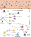
Figure 2
Immune cells in brain after ischemic stroke. Cerebral ischemic stroke leads to the release of danger-associated molecular patterns (DAMPs) from dying neurons. These molecules trigger the activation of resident microglia and astrocytes. Activated microglia release pro- or anti-inflammatory mediators resulting in neurons apoptosis or neurogenesis. In the ischemic brain, inflamed endothelial cells express adhesion molecules, such as intercellular adhesion molecule 1 (ICAM-1), P-selectin, and E-selectin, which recruit neutrophils and infiltration. Activated neutrophils infiltrating into the brain parenchyma produce a number of inflammatory factors, including matrix metalloproteinases (MMPs), inducible nitric oxide synthase (iNOS), and reactive oxygen species (ROS), which aggravate blood brain barrier destruction and cell death, as well as obstruct brain repair. Dendritic cells express major histocompatibility complex (MHC II) on the cell surface. Brain-derived antigens can be presented by MHC II on dendritic cell and can be recognized by receptors on the surface of T cells. Subsequently, the adaptive immune system is activated. Activated microglia/macrophages may stimulate activated CD4+T cells to differentiate into Th1 or Th2 cells, and then produce pro-inflammatory or anti-inflammatory cytokines to damage or protect the brain. CD8+ cytotoxic T cells lead to neuron death and aggravation of brain injury by cell interactions and release of perforin/granzyme after antigen-dependent activation. γδT cells caused damage to the surrounding tissues by secreting IL-17. B cells attenuate inflammation after stroke by producing anti-inflammatory cytokines such as IL-10 and TGF-β.
Figure 3
Figure 3
Different cell subsets changes in response to stroke. Two main subgroups of monocytes exist in the circulation, namely pro-inflammatory Ly6ChiCCR2+CX3CR1lo monocytes and anti-inflammatory Ly6CloCCR2CX3CR1hi monocytes. Ly6ChiCCR2+CX3CR1lo monocytes infiltrate into the central nervous system from the blood via the CCL2-CCR2 axis, and differentiate into classically M1-like macrophages or Tip-DCs with strong phagocytosis. Under acute inflammatory circumstances, they turn into the direct precursors of macrophages in the peripheral blood. Anti-inflammatory monocytes are larger and act primarily as vascular patrols and induce neutrophil aggregation. After cerebral ischemia, the injury tissue releases various inflammatory cytokines. Lipopolysaccharide (LPS) and interferon-γ (IFN-γ) stimulate monocyte-derived macrophages to polarize toward M1 phenotype which secretes TNF-α, IL-1β, and IL-6. Alternative M2 is promoted by IL-4, IL-10, and TGF-β. It expresses substantial mannose receptors and scavenger receptors.

References

    1. Sternberg Z, Schaller B. Central noradrenergic agonists in the treatment of ischemic stroke-an overview. Transl Stroke Res. (2019) 10.1007/s12975-019-00718-7. [Epub ahead of print]. - DOI - PubMed
    1. Bayraktutan U. Endothelial progenitor cells: Potential novel therapeutics for ischaemic stroke. Pharmacol Res. (2019) 144:181–91. 10.1016/j.phrs.2019.04.017 - DOI - PubMed
    1. Petrovic-Djergovic D, Goonewardena SN, Pinsky DJ. Inflammatory disequilibrium in stroke. Circ Res. (2016) 119:142–58. 10.1161/CIRCRESAHA.116.308022 - DOI - PMC - PubMed
    1. Anrather J, Iadecola C. Inflammation and stroke: an overview. Neurotherapeutics. (2016) 13:661–70. 10.1007/s13311-016-0483-x - DOI - PMC - PubMed
    1. Leech T, Chattipakorn N, Chattipakorn SC. The beneficial roles of metformin on the brain with cerebral ischaemia/reperfusion injury. Pharmacol Res. (2019) 146:104261. 10.1016/j.phrs.2019.104261 - DOI - PubMed

Publication types