. 2019 Oct;18(4):2628-2638.
doi: 10.3892/etm.2019.7863. Epub 2019 Aug 7.

Gene delivery into hepatic cells with ternary complexes of plasmid DNA, cationic liposomes and apolipoprotein E-derived peptide

Affiliations

Gene delivery into hepatic cells with ternary complexes of plasmid DNA, cationic liposomes and apolipoprotein E-derived peptide

Yoshiyuki Hattori et al. Exp Ther Med. 2019 Oct.

Abstract

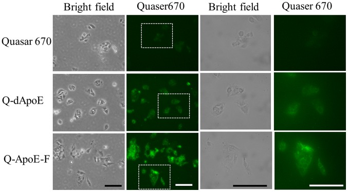
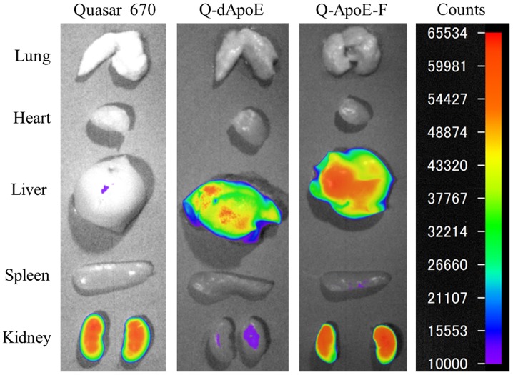
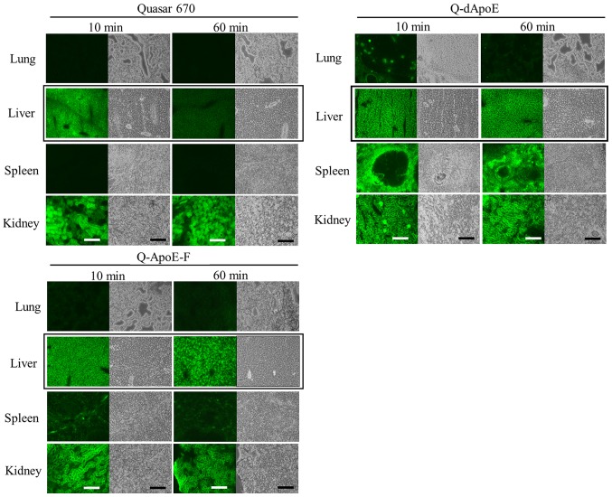
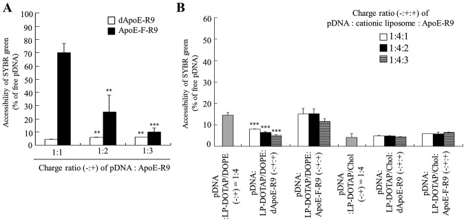
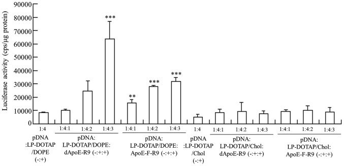
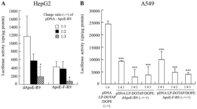
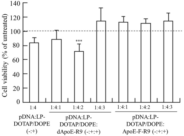
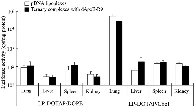
Cationic liposomes containing a cationic lipid, such as 1,2-dioleoyl-3-trimethylammonium-propane (DOTAP), have often been used for the transduction of plasmid DNA (pDNA) in vivo. However, such liposomes induce gene expression primarily in the lungs after intravenous injection. To improve the delivery of cationic liposomes/pDNA complexes (pDNA lipoplexes) to the liver by intravenous administration, the current study synthesized two apolipoprotein E (ApoE)-derived peptides, dApoE-R9 and ApoE-F-R9, for liver targeting via certain ApoE receptors, including the low-density lipoprotein receptor. Ternary complexes of pDNA, cationic liposomes and ApoE-R9 peptide were also prepared. After in vitro transfection, ternary complexes with DOTAP/1,2-dioleoyl-sn-glycero-3-phosphoethanolamine (DOPE) liposomes exhibited high transfection activity in HepG2 cells compared with DOTAP/cholesterol (Chol) liposomes. In particular, ternary complexes with dApoE-R9 exhibited high transfection activity in cells compared with ApoE-F-R9. However, in vivo transfection studies revealed that ternary complexes with DOTAP/DOPE liposomes and dApoE-R9 did not increase gene expression in the liver compared with DOTAP/DOPE lipoplexes. In contrast, ternary complexes with DOTAP/Chol liposomes and dApoE-R9 increased gene expression in the liver compared with DOTAP/Chol lipoplexes. The results demonstrated that the in vivo optimal liposomal formulation in ternary complexes with ApoE-R9 peptide for liver delivery were different from those that were in vitro.

Keywords: apolipoprotein E; cationic liposome; liver-targeting; plasmid DNA.


Figures

Figure 1.
Figure 1.
Intracellular localization of ApoE-derived peptide in HepG2 cells after incubation for 3 h. Free Quaser670, Q-dApoE, and Q-ApoE-F were diluted in 1 ml culture medium at final concentrations of 1, 10 and 10 µg/ml (2, 2.8 and 2.6 µM), respectively, and then incubated with cells for 3 h. Green signals indicate localization of Quaser670, Q-dApoE or Q-ApoE-F. White boxes indicate the enlarged panels presented in the right panels. Scale bar, 100 µm. ApoE, apolipoprotein E.
Figure 2.
Figure 2.
Biodistribution of ApoE-derived peptide in mice 1 h after intravenous injection. Q-dApoE or Q-ApoE-F (20 µg) was administered intravenously to mice. As a control, Quaser670 (2 µg) was administered intravenously. A total of 1 h after injection, mice were sacrificed and Quaser670 fluorescence imaging of the tissues was performed. Fluorescence intensity is illustrated by a color-coded scale (red is maximum, purple is minimum). ApoE, apolipoprotein E.
Figure 3.
Figure 3.
Biodistribution of ApoE-derived peptide in mice at 10 and 60 min after intravenous injection. Q-dApoE or Q-ApoE-F (100 µg) was administered intravenously to mice. As a control, Quaser670 (10 µg) was administered intravenously. A total of 10 or 60 min after injection, mice were sacrificed and tissues were frozen on dry ice and sliced into 16 µm sections. The localization of Quaser670 was examined using a fluorescence microscope. Green signals indicated the localization of Quaser670, Q-dApoE or Q-ApoE-F. Black boxes indicate marked changes of accumulation in the liver. Scale bar, 200 µm. ApoE, apolipoprotein E.
Figure 4.
Figure 4.
Association of pDNA with ApoE-derived peptide and cationic liposomes as determined via an exclusion assay using SYBR® Green I Nucleic Acid Gel Stain. (A) Binary complexes of pDNA and ApoE-R9 peptide were formed at various charge ratios (−:+) from 1:1-1:3. **P<0.01 and ***P<0.001 vs. the binary complexes of dApoE-R9 or ApoE-F-R9 at a charge ratio (−:+) of 1:1. (B) pDNA lipoplexes were prepared by mixing pDNA with cationic liposomes (LP-DOTAP/DOPE or LP-DOTAP/Chol) at a charge ratio (−:+) of 1:4. Ternary complexes of pDNA, cationic liposomes and ApoE-R9 peptide were formed at various charge ratios (−:+:+) from 1:4:1 to 1:4:3. As a control, the value of fluorescence obtained after the addition of free pDNA solution was set as 100%. The quantity of pDNA available to interact with the SYBR® Green I is expressed as a percentage of the control. ***P<0.001 vs. the LP-DOTAP/DOPE lipoplexes. Data are presented as the mean + standard deviation (n=3). pDNA, plasmid DNA; ApoE, apolipoprotein E; LP, liposome; DOTAP, 1,2-dioleoyl-3-trimethylammonium-propane methyl sulfate salt; DOPE, 1,2-dioleoyl-sn-glycero-3-phosphoethanolamine.
Figure 5.
Figure 5.
Effect of charge ratio (−:+:+) of pDNA, cationic liposome and ApoE-R9 peptide on the luciferase activity of HepG2 cells at 24 h after transfection with the ternary complex. pDNA lipoplexes were prepared by mixing pCMV-Luc with LP-DOTAP/Chol or LP-DOTAP/DOPE at a charge ratio (−:+) of 1:4. Ternary complexes of pCMV-Luc, cationic liposome and ApoE-R9 peptide were prepared at charge ratios (−:+:+) from 1:4:1-1:4:3. Data are presented as the mean + standard deviation (n=3). **P<0.01 and ***P<0.001 vs. LP-DOTAP/DOPE lipoplexes. pDNA, plasmid DNA; ApoE, apolipoprotein E; LP, liposome; DOTAP, 1,2-dioleoyl-3-trimethylammonium-propane methyl sulfate salt; Chol, cholesterol; DOPE, 1,2-dioleoyl-sn-glycero-3-phosphoethanolamine.
Figure 6.
Figure 6.
Luciferase activity in HepG2 cells 24 h after transfection with binary complexes of pDNA and ApoE-R9 peptide, and in A549 cells 24 h after transfection with ternary complexes. (A) binary complexes were prepared by mixing pCMV-Luc with dApoE-R9 or ApoE-F-R9 at charge ratios (−:+) from 1:1-1:3. *P<0.05 vs. binary complexes of dApoE-R9 or ApoE-F-R9 at a charge ratio (−:+) of 1:1. (B) pDNA lipoplexes were prepared by mixing pCMV-Luc with cationic liposomes (LP-DOTAP/DOPE or LP-DOTAP/Chol) at a charge ratio (−:+) of 1:4. Ternary complexes of pCMV-Luc, cationic liposome, and ApoE-R9 peptide were prepared at charge ratios (−:+:+) from 1:4:1-1:4:3. ***P<0.001 vs. LP-DOTAP/DOPE lipoplexes. Data are presented as the mean + standard deviation (n=3). pDNA, plasmid DNA; ApoE, apolipoprotein E; LP, liposome; DOTAP, 1,2-dioleoyl-3-trimethylammonium-propane methyl sulfate salt; DOPE, 1,2-dioleoyl-sn-glycero-3-phosphoethanolamine; Chol, cholesterol.
Figure 7.
Figure 7.
Cell viability 24 h after transfection with pDNA lipoplexes and ternary complexes in HepG2 cells. pDNA lipoplexes were prepared by mixing pDNA with LP-DOTAP/DOPE at a charge ratio (−:+) of 1:4 and ternary complexes were prepared by mixing pDNA with LP-DOTAP/DOPE and the ApoE-R9 peptide at charge ratios (−:+:+) from 1:4:1-1:4:3. Data are presented as the mean + standard deviation (n=4-6). ***P<0.001 vs. untreated cells. pDNA, plasmid DNA; LP, liposome; DOTAP, 1,2-dioleoyl-3-trimethylammonium-propane methyl sulfate salt; DOPE, 1,2-dioleoyl-sn-glycero-3-phosphoethanolamine; ApoE, apolipoprotein E.
Figure 8.
Figure 8.
Luciferase activities at 24 h after intravenous administration of pDNA lipoplexes and ternary complexes into mice. pDNA lipoplexes were prepared by mixing pCMV-Luc with cationic liposomes (LP-DOTAP/DOPE or LP-DOTAP/Chol) at a charge ratio (−:+) of 1:4. Ternary complexes were prepared by mixing pCMV-Luc with cationic liposomes and dApoE-R9 at a charge ratio (−:+:+) of 1:4:3. The pDNA lipoplexes or ternary complexes with 30 µg of pCMV-Luc were administered intravenously via the lateral tail vein into mice. The difference of luciferase activity between pDNA lipoplexes and ternary complexes with dApoE-R9 was not statistically significant in any tissue. Data are presented as the mean + standard deviation (n=3). pDNA, plasmid DNA; LP, liposome; DOTAP, 1,2-dioleoyl-3-trimethylammonium-propane methyl sulfate salt; DOPE, 1,2-dioleoyl-sn-glycero-3-phosphoethanolamine; Chol, cholesterol; ApoE, apolipoprotein E.

Similar articles

Cited by

References

    1. Aravalli RN, Belcher JD, Steer CJ. Liver-targeted gene therapy: Approaches and challenges. Liver Transpl. 2015;21:718–737. doi: 10.1002/lt.24122. - DOI - PMC - PubMed
    1. Wu J, Nantz MH, Zern MA. Targeting hepatocytes for drug and gene delivery: Emerging novel approaches and applications. Front Biosci. 2002;7:d717–d725. doi: 10.2741/A806. - DOI - PubMed
    1. Nayerossadat N, Maedeh T, Ali PA. Viral and nonviral delivery systems for gene delivery. Adv Biomed Res. 2012;1:27. doi: 10.4103/2277-9175.98152. - DOI - PMC - PubMed
    1. Palaschak B, Herzog RW, Markusic DM. AAV-mediated gene delivery to the liver: Overview of current technologies and methods. Methods Mol Biol 1950. 2019:333–360. doi: 10.1007/978-1-4939-9139-6_20. - DOI - PubMed
    1. Zhang Y, Bradshaw-Pierce EL, Delille A, Gustafson DL, Anchordoquy TJ. In vivo comparative study of lipid/DNA complexes with different in vitro serum stability: Effects on biodistribution and tumor accumulation. J Pharm Sci. 2008;97:237–250. doi: 10.1002/jps.21076. - DOI - PubMed