. 2019 Sep 25;5(9):1514-1522.
doi: 10.1021/acscentsci.9b00416. Epub 2019 Aug 19.

PhoX: An IMAC-Enrichable Cross-Linking Reagent

Affiliations

PhoX: An IMAC-Enrichable Cross-Linking Reagent

Barbara Steigenberger et al. ACS Cent Sci. .

Abstract

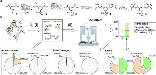
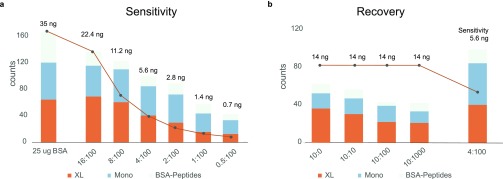
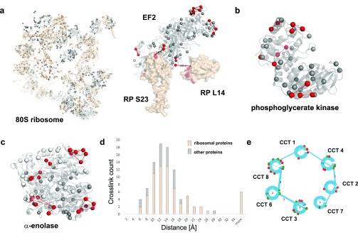
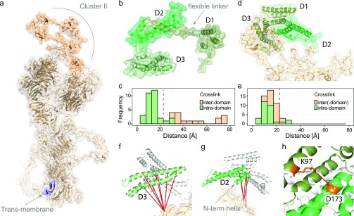
Chemical cross-linking mass spectrometry is rapidly emerging as a prominent technique to study protein structures. Structural information is obtained by covalently connecting peptides in close proximity by small reagents and identifying the resulting peptide pairs by mass spectrometry. However, substoichiometric reaction efficiencies render routine detection of cross-linked peptides problematic. Here, we present a new trifunctional cross-linking reagent, termed PhoX, which is decorated with a stable phosphonic acid handle. This makes the cross-linked peptides amenable to the well-established immobilized metal affinity chromatography (IMAC) enrichment. The handle allows for 300× enrichment efficiency and 97% specificity. We exemplify the approach on various model proteins and protein complexes, e.g., resulting in a structural model of the LRP1/RAP complex. Almost completely removing linear peptides allows PhoX, although noncleavable, to be applied to complex lysates. Focusing the database search to the 1400 most abundant proteins, we were able to identify 1156 cross-links in a single 3 h measurement.


Conflict of interest statement

The authors declare no competing financial interest.

Figures

Figure 1
Figure 1
PhoX synthesis, workflow, and performance. (a) The synthesis of PhoX can be achieved in five steps starting from commercially available precursors. (b) After cross-linking the proteins in their native state, the proteins are denatured, reduced, alkylated, and digested to peptides (I). The mixture of peptides contains 3 different kinds of products: unmodified peptides (gray), monolinks (orange), and cross-links (green) (II); these colors are reused. Cross-linked and monolinked peptides are enriched using Fe-IMAC on a liquid sample handling platform providing high sample throughput (III; used data is from the BSA experiment presented in the Supporting Information, Note 8). Direct measurement of the cross-linked peptides produces low counts of cross-link identifications due to their extremely low abundances (left panel), while measurement following Fe-IMAC enrichment results in no cross-link identifications in the flow-through with similar abundance levels for the linear peptides as detected in the No enrichment (middle panel) and many in the eluate due to their enhanced abundance levels (right panel).
Figure 2
Figure 2
Testing PhoX peptide enrichment in a complex background. (a) Sensitivity—mixing cross-linked BSA peptides in decreasing amounts in a fixed background of E. coli peptides provides an estimate of the detection limit of the methodology. (b) Recovery method—mixing cross-linked BSA peptides in equal amounts in an increasing background of E. coli peptides allows for recovery of cross-linked BSA peptides upon enrichment. The estimated cross-link amounts for the given cross-linked BSA peptides are shown above the bars.
Figure 3
Figure 3
Application of PhoX to investigate human cell lysates. Cross-linked lysines are depicted as red spheres; non-cross-linked lysines are depicted as gray spheres. (a) Ribosome cryo-EM map (PDB, 4v6x). Proteins found cross-linked are shown in light brown; proteins without cross-links are shown in gray (RNA is not shown). Intra-cross-links mapped on the elongation factor 2 (part of PDB, 4v6x). Inter-cross-links of elongation factor to ribosomal proteins mapped on the ribosome (PDB, 6v6x). (b) Intra-cross-links of phosphoglycerate kinase mapped on its crystal structure (PDB, 3c39) (c) Intra-cross-links of α-enolase mapped on the dimeric crystal structure of human enolase 1 (PDB, 3b97). (d) Histogram of observed Lys–Lys distances in PhoX cross-links on the 80S ribosome (colored in light brown) and other proteins (nucleolin, PDB, 2kkr; nucleophosmin, PDB, 2llh; elongation factor 2, PDB, 4v6x; stress-induced-phosphoprotein 1, PDB, 1ewl; α-enolase, PDB, 3b97; phosphoglycerate kinase, PDB, 3c39). (e) Interaction network found for the TRiC/CCT complex. Proteins of the TRiC/CCT complex are shown as blue circles. Interlinks between the proteins are shown as blue lines; the thickness of the lines indicates the amount of interlinks identified. Green lines in the circles are the lysine positions; red balls indicate phosphorylation sites as extracted from uniprot, and green balls indicate lysines involved in interlinks.
Figure 4
Figure 4
Application of PhoX to investigate the binding of full-length RAP to LRP1 Cluster II. (a) Predicted structural model for full-length LRP1, with Cluster II (PDB, 1n7d) and the transmembrane region highlighted. (b) Structure of RAP with the individual domains indicated (predicted from PDB, 2p03). (c) Measured Cα–Cα distances for intra- and interdomain cross-links on the structure of RAP (maximum distance constraint indicated with red, dashed line). (d) Multibody docking of the individual domains of RAP to LRP1 using HADDOCK. (e) Measured Cα–Cα distances after docking (maximum distance constraint indicated with red, dashed line). (f) Cross-links between the D3 domain of RAP and LRP1. (g) Cross-links between the D2 domain of RAP and LRP1. (h) Domain D1 (K97) in close proximity to domain D2 (D173).

References

    1. Leitner A.; Faini M.; Stengel F.; Aebersold R. Crosslinking and Mass Spectrometry: An Integrated Technology to Understand the Structure and Function of Molecular Machines. Trends Biochem. Sci. 2016, 41 (1), 20–32. 10.1016/j.tibs.2015.10.008. - DOI - PubMed
    1. Holding A. N. XL-MS: Protein Cross-Linking Coupled with Mass Spectrometry. Methods 2015, 89, 54–63. 10.1016/j.ymeth.2015.06.010. - DOI - PubMed
    1. Rappsilber J. The Beginning of a Beautiful Friendship: Cross-Linking/Mass Spectrometry and Modelling of Proteins and Multi-Protein Complexes. J. Struct. Biol. 2011, 173 (3), 530–540. 10.1016/j.jsb.2010.10.014. - DOI - PMC - PubMed
    1. Merkley E. D.; Cort J. R.; Adkins J. N. Cross-Linking and Mass Spectrometry Methodologies to Facilitate Structural Biology: Finding a Path through the Maze. J. Struct. Funct. Genomics 2013, 14 (3), 77–90. 10.1007/s10969-013-9160-z. - DOI - PMC - PubMed
    1. Sinz A. Chemical Cross-Linking and Mass Spectrometry to Map Three-Dimensional Protein Structures and Protein–Protein Interactions. Mass Spectrom. Rev. 2006, 25 (4), 663–682. 10.1002/mas.20082. - DOI - PubMed