

Mouse (Mus Musculus) Embryonic Cerebral Cortex Cell Death Caused by Carbofuran Insecticide Exposure
- PMID: 31572823
- PMCID: PMC6749746
- DOI: 10.2478/jvetres-2019-0040
Mouse (Mus Musculus) Embryonic Cerebral Cortex Cell Death Caused by Carbofuran Insecticide Exposure
Abstract
Introduction: The aim of the study was to describe the process of neuron death in the cerebral cortex caused by embryonic carbofuran exposure.
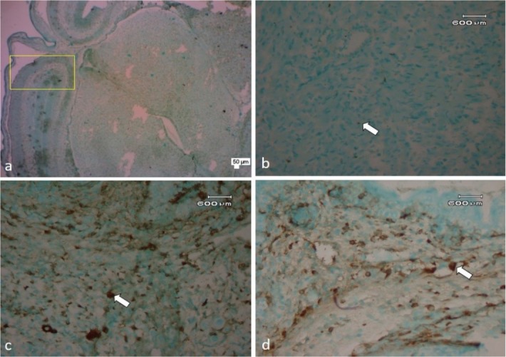
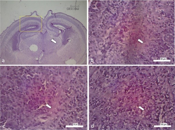
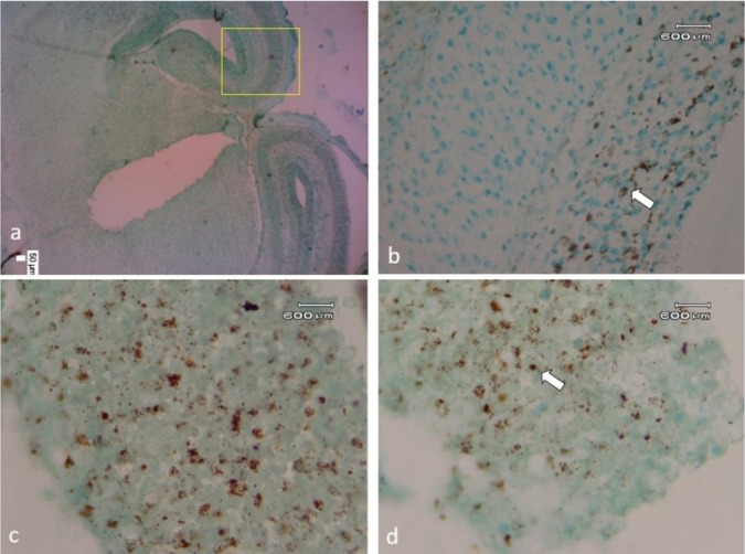
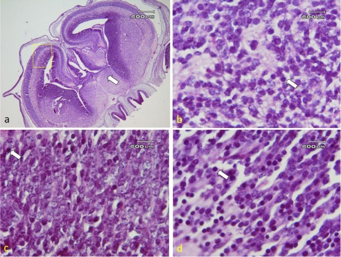
Material and methods: 81 mouse foetuses from 27 breeding mice were used in the study. Carbofuran was administered by gavage from the 6th to the 15th day of gestation to two groups: one at 0.0208 and the other at 0.0417 mg/kg b.w. On the 17th day, the mice were sacrificed and the foetuses were taken to measure the ROS (malondialdehyde/MDA and superoxide dismutase/SOD) activity in brain tissue, the number of apoptotic embryonic cerebral cortex neurons using a TUNEL assay, and necrotic cells using HE staining. Examination of p53 and caspase 3 expression was done by immunohistochemistry. Data were analysed using analysis of variance (ANOVA) and Duncan's test.
Results: Increased activity of cerebral ROS characterised by significant elevation of the MDA level (P < 0.05), decreased SOD (P < 0.01), increased p53 and caspase 3 expression, and cerebral cortical neuron death either by necrosis or apoptosis (P < 0.05) were found. At the low dose carbofuran increased expression of p53, caspase 3, and apoptosis. At the high dose it increased levels of MDA and necrosis.
Conclusion: Increased expression of p53 and caspase 3 and apoptosis indicated that carbofuran may cause apoptosis through the intrinsic pathway. The increased apoptosis grants an opportunity to prevent and treat the effect of ROS due to gestational carbofuran exposure.
Keywords: ROS; apoptosis; carbofuran; mice; necrosis.
© 2019 E.M. Luqman et al. published by Sciendo.
Conflict of interest statement
Conflict of Interest Conflict of Interests Statement: The authors declare that there is no conflict of interests regarding the publication of this article.
Figures
References
-
- Anna K., Bal-Price J., Helena T.H. Stoytcheva M. Pesticides - The impacts of pesticide exposure. InTech; London: 2011. Effects of pesticides on neuronal and glial cell differentiation and maturation in primary cultures; pp. 341–356. Edited by.
-
- Bolaris S., Bozas E., Benekou A., Phillippiis H., Stylianopoulou F.. In utero radiation-induced apoptosis and p53 gene expression in the developing rat brain. Int J Radiat Biol. 2001;77:71–81. - PubMed
-
- Chandra D., Tripathi U.N., Srivastava S., Swaroop A.. Carbofuran induced biochemical toxicity in mice: Protective role of Momordica charantia. Euro J Exp Bio. 2011;1:106–112.
-
- Conti M., Morand P.C., Levillain P., Lemonnier A.. Improved fluorometric determination of malonaldehyde. Clin Chem. 1991;37:1273–125. - PubMed
-
- Ghezi P., Brines M.. Erythropoietin as an antiapoptotic, tissue-protective cytokine. Cell Death Differ. 2004;11(1):37–44. - PubMed
LinkOut - more resources
Full Text Sources
Research Materials
Miscellaneous