. 2019 Dec;44(6):2091-2102.
doi: 10.3892/ijmm.2019.4359. Epub 2019 Oct 1.

Protective effect of hydrogen sulfide on monocrotaline‑induced pulmonary arterial hypertension via inhibition of the endothelial mesenchymal transition

Affiliations

Protective effect of hydrogen sulfide on monocrotaline‑induced pulmonary arterial hypertension via inhibition of the endothelial mesenchymal transition

Hui Zhang et al. Int J Mol Med. 2019 Dec.

Abstract

Endothelial‑to‑mesenchymal transition (EndMT) serves an important role in the vascular remodeling of pulmonary arterial hypertension (PAH). However, little is known about the correlation between hydrogen sulfide (H2S), a protective gaseous mediator in PAH and the process of EndMT. Male Sprague‑Dawley rats (10 weeks old) received a single dose of monocrotaline (MCT; i.p., 60 mg/kg) and were randomly treated with NaHS [an H2S donor; intraperitoneal (i.p.) 1 mg/kg/day], DL‑propagylglycine (an inhibitor of H2S synthesis; PAG; i.p., 10 mg/kg/day) or saline, 7 days after MCT injection. Rats were sacrificed 21 days after MCT injection. A selection of human pulmonary artery endothelial cells (HPAECs) were pretreated with NaHS or saline and stimulated with transforming growth factor (TGF)‑β1 (10 ng/ml), and the other HPAECs were transfected with a cystathionine γ‑lyase (CSE, an H2S synthesizing enzyme) plasmid and subsequently stimulated with TGF‑β1. NaHS was indicated to inhibit EndMT and PAH progression by inhibiting the induction of the nuclear factor (NF)‑κB‑Snail pathway. In contrast, the depletion of H2S formation by PAG exacerbated EndMT and PAH by activating NF‑κB‑Snail molecules. In HPAECs, NaHS dose‑dependently inhibited TGF‑β1‑induced EndMT and the activation of the NF‑κB‑Snail pathway. Transfection with a CSE plasmid significantly repressed TGF‑β1‑induced expression of the mesenchymal marker and upregulated the expression of the endothelial marker, which was accompanied by the suppression of the NF‑κB‑Snail pathway. The inhibitory effect of CSE overexpression on TGF‑β1‑induced EndMT was significantly reversed by pretreatment with PAG. In conclusion, the current study provides novel information elucidating the beneficial effect of H2S on PAH through inhibiting the induction of the NF‑κB‑Snail pathway and the subsequent process of EndMT in pulmonary arteries.


Figures

Figure 1
Figure 1
Time dependent alterations in protein signatures of EndMT and the dynamic characteristics of endogenous H2S synthesis in MCT-induced PAH. After injection with MCT, rats were sacrificed at days 0, 7, 14, 21 and 28. Temporal characteristics of VE-cadherin and α-SMA in lungs were measured using (A) western blot analysis and (B) immunofluorescent staining. Red fluorescence represents VE-cadherin, green fluorescence represents α-SMA and blue fluorescence indicates DAPI nuclei staining. Expression of (A) Snail and (C) CSE in the lungs was measured using western blot analysis. (D) Lung CSE enzymatic activity and (E) plasma H2S level were also measured. Results are presented as the mean ± standard error of the mean (n=6 animals in each group). *P<0.05 vs. rats at baseline; #P<0.05 vs. rats 7 days after MCT injection; &P<0.05 vs. rats 14 days after MCT injection. EndMT, endothelial-to-mesenchymal transition; H2S, hydrogen sulfide; MCT, monocrotaline; PAH, pulmonary arterial hypertension; CSE, cystathionine γ-lyase; SMA, smooth muscle actin; VE, vascular endothelial; DAPI, 4,6-diamidino-2-phenylindole.
Figure 2
Figure 2
Effect of supplementation with exogenous H2S (NaHS) or inhibition of H2S formation by PAG on MCT-induced PAH. Rats received NaHS (i.p., 1 mg/kg/day) or PAG (i.p, 10 mg/kg/day) 7 days after MCT injection. A period of 21 days after injection of MCT, rats were anesthetized and underwent RHC. (A) Pulmonary artery systolic pressure, pulmonary arterial diastolic pressure and pulmonary arterial mean pressure were continuously recorded. (B) The RV/(LV+S) ratio was calculated, as described in materials and methods, to estimate RV hypertrophy. (C) Right ventricular ejection fraction was indicated by TAPSE measured using ultrasound and (D) quantitative analysis. (E) Pulmonary vascular remodeling was assessed using hematoxylin and eosin staining. (F) Media to lumen ratio (MA/LA) was measured as described in the materials and methods, to evaluate the remodeling of pulmonary arteries. The results are presented as the mean ± standard error of the mean (n=6 animals in each group). *P<0.05 vs. the control group; #P<0.05 vs. rats with MCT+saline. H2S, hydrogen sulfide; PAG, DL-propagylglycine; PAH, pulmonary arterial hypertension; RV/(LV+S), weight ratio of the right ventricle to left ventricle plus septum; MCT, monocrotaline. TAPSE, tricuspid annular plane systolic excursion. MA, media area; LA, lumen area; i.p., intraperitoneal.
Figure 3
Figure 3
Effect of NaHS or PAG on EndMT in MCT-induced PAH. Rats received NaHS (i.p., 1 mg/kg/day) or PAG (i.p, 10 mg/kg/day) 7 days after MCT injection. A period of 21 days after injection of MCT, rats were sacrificed (n=6 animals in each group). The pulmonary expression levels of EndMT markers, VE-cadherin and α-SMA were examined using (A) western blot analysis and (B) immunofluorescent staining. Red fluorescence represents VE-cadherin, green fluorescence represents α-SMA and blue fluorescence represents 4,6-diamidino-2-phenylindole nuclei staining. (C) The ultrastructure of the pulmonary artery was assessed using transmission electron microscopy. In the control group, endothelial cells are flat, elongated and separated from smooth muscle cells via a thick basement membrane, the BL. In the MCT group, endothelial cells display migration in the intima and alter their nuclei orientation. Endothelial characteristics of these cells are confirmed by the presence of caveolae (black arrow) and Weibel-Palade bodies (white arrows). *P<0.05 vs. the control group; #P<0.05 vs. rats with MCT+saline. NaHS, exogenous H2S; PAG, DL-propagylglycine; PAH, pulmonary arterial hypertension; EndMT, endothelial-to-mesenchymal transition; MCT, monocrotaline; EC, endothelial cells; SMC, smooth muscle cells; BL, basal lamina; i.p., intraperitoneal; SMA, smooth muscle actin; VE, vascular endothelial.
Figure 4
Figure 4
Effect of NaHS or PAG on NF-κB-Snail pathway in MCT-induced PAH. Rats received NaHS (i.p., 1 mg/kg/day) or PAG (i.p, 10 mg/kg/day) 7 days after MCT injection. A period of 21 days after injection of MCT, rats were sacrificed. Pulmonary expression of Snail, phospho-IκBα and IκBα were measured using (A) western blot analysis. (B) p65 DNA binding activity in lung nuclear extracts were measured as described in the materials and methods. The results are presented as the mean ± standard error of the mean (n=6 animals in each group). *P<0.05 vs. the control group; #P<0.05 vs. rats with MCT+saline. NaHS, exogenous H2S; PAG, DL-propagylglycine; PAH, pulmonary arterial hypertension; MCT, monocrotaline; phospho, phosphorylated; i.p., intraperitoneal; NF, nuclear factor.
Figure 5
Figure 5
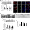
Effect of NaHS on TGF-β1-induced EndMT in HPAECs. HPAECs were pre-incubated with saline or NaHS (50, 100 and 200 µM) for 2 h and stimulated with TGF-β1 (10 ng/ml) for 24 h in the continuous presence of NaHS (50, 100 and 200 µM) or saline. Some cells were harvested to investigate the changes in NF-κB-Snail pathway. Remaining cells were subsequently stimulated with TGF-β1 (10 ng/ml) for an additional 9 days in an attempt to estimate the process of EndMT. Expression of EndMT markers, VE-cadherin and α-SMA was examined using (A) western blot analysis and (B) immunofluorescent staining. Red fluorescence represents VE-cadherin, green fluorescence represents α-SMA and blue fluorescence represents DAPI nuclei staining. The ultrastructure of HPAECs was assessed using (C) TEM. Black arrows indicate caveolae. White arrows indicate WPB. Black triangles indicate microfilaments in the cytoplasm. Effect of NaHS on cell morphology in TGF-β1-induced EndMT in HPAECs was investigated using (D) phase-contrast light microscopy. Normal HPAECs exhibit a cobblestone appearance and indicate caveolae and WPB, with few microfilaments. Following exposure to TGF-β1, microfilamentation appeared in the cytoplasm and cells are elongated and spindle-shaped. The expression of Snail, phospho-IκBα and IκBα was measured using (E) western blot analysis. (F) p65 DNA binding activity in nuclear extracts was measured as described in the materials and methods. The data are presented as the mean ± standard error of the mean of at least three independent experiments. *P<0.05 vs. the control group at baseline; #P<0.05 vs. TGF-β1-stimulated HPAECs treated with saline; &P<0.05 vs. TGF-β1-stimulated HPAECs treated with NaHS at a concentration of 50 µM. NaHS, exogenous H2S; EndMT, endothelial-to-mesenchymal transition; HPAEC, human pulmonary artery endothelial cells; WPB, Weibel-Palade bodies; TEM, transmission electron microscopy; VE, vascular endothelial; TGF, transforming growth factor; DAPI, 4,6-diamidino-2-phenylindole.
Figure 6
Figure 6
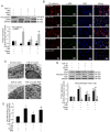
Effect of CSE overexpression on TGF-β1-induced EndMT in HPAECs. HPAECs were transfected with a vector or CSE plasmid and stimulated with TGF-β1 (10 ng/ml) for 24 h in the presence of PAG (2 mM) or saline. These cells were harvested to investigate the changes in the NF-κB-Snail pathway. Some cells were subsequently stimulated with TGF-β1 (10 ng/ml) for an additional 9 days to estimate the process of EndMT. Expression of EndMT markers, VE-cadherin and α-SMA and signal molecules were examined using (A) western blotting and (B) immunofluorescent staining. Red fluorescence represents VE-cadherin, green fluorescence represents α-SMA and blue fluorescence represents DAPI nuclei staining. Scale bar=50 µm. (C) The ultrastructure of HPAECs was assessed using TEM. White arrows indicate caveolae. Black arrows indicate WPB. Black triangles indicate microfilaments in the cytoplasm. (D) Expression of Snail, phospho-IκBα and IκBα were measured using western blot analysis. (E) p65 DNA binding activity in nuclear extracts were measured as described in the materials and methods. The data are presented as the mean ± standard error of the mean of at least three independent experiments. *P<0.05 vs. control group; #P<0.05 vs. TGF-β1-stimulated HPAECs transfected with vector; &P<0.05 vs. TGF-β1-stimulated HPAECs transfected with CSE plasmid. CSE, cystathionine γ-lyase; EndMT, endothelial-to-mesenchymal transition; DAPI, 4,6-diamidino-2-phenylindole; HPAEC, human pulmonary artery endothelial cells; PAG, DL-propagylglycine; WPB, Weibel-Palade bodies; TEM, transmission electron microscopy; TGF, transforming growth factor; NF, nuclear factor; phospho, phosphorylated; VE, vascular endothelial.

Similar articles

Cited by

References

    1. Goumans MJ, van Zonneveld AJ, ten Dijke P. Transforming growth factor beta-induced endothelial-to-mesenchymal transition: A switch to cardiacfibrosis? Trends Cardiovasc Med. 2008;18:293–298. doi: 10.1016/j.tcm.2009.01.001. - DOI - PubMed
    1. Willis BC, Borok Z. TGF-beta-induced EMT: Mechanisms and implications for fibrotic lung disease. Am J Physiol Lung Cell Mol Physiol. 2007;293:L525–L534. doi: 10.1152/ajplung.00163.2007. - DOI - PubMed
    1. Zeisberg EM, Potenta SE, Sugimoto H, Zeisberg M, Kalluri R. Fibroblasts in kidney fibrosis emerge via endothelial-to-mesenchymal transition. J Am Soc Nephrol. 2008;19:2282–2287. doi: 10.1681/ASN.2008050513. - DOI - PMC - PubMed
    1. Zeisberg EM, Tarnavski O, Zeisberg M, Dorfman AL, McMullen JR, Gustafsson E, Chandraker A, Yuan X, Pu WT, Roberts AB, et al. Endothelial-to-mesenchymal transition contributes to cardiac fibrosis. Nat Med. 2007;13:952–961. doi: 10.1038/nm1613. - DOI - PubMed
    1. Arciniegas E, Frid MG, Douglas IS, Stenmark KR. Perspectives onendothelial-to-mesenchymal transition: Potential contribution to vascular remodeling in chronic pulmonary hypertension. Am J Physiol Lung Cell Mol Physiol. 2007;293:L1–L8. doi: 10.1152/ajplung.00378.2006. - DOI - PubMed

MeSH terms