. 2019 Nov;44(5):1943-1951.
doi: 10.3892/ijmm.2019.4351. Epub 2019 Sep 26.

S‑allyl‑cysteine sulfoxide (alliin) alleviates myocardial infarction by modulating cardiomyocyte necroptosis and autophagy

Affiliations

S‑allyl‑cysteine sulfoxide (alliin) alleviates myocardial infarction by modulating cardiomyocyte necroptosis and autophagy

Li-Jun Yue et al. Int J Mol Med. 2019 Nov.

Abstract

S‑allyl‑cysteine sulfoxide (alliin) is the main organosulfur component of garlic and its preparations. The present study aimed to examine the protective effect of alliin on cardiac function and the underlying mechanism in a mouse model of myocardial infarction (MI). Notably, alliin treatment preserved heart function, attenuated the area of infarction in the myocardium of mice and reduced lesions in the myocardium, including cardiomyocyte fibrosis and death. Further mechanistic experiments revealed that alliin inhibited necroptosis but promoted autophagy in vitro and in vivo. Cell viability assays showed that alliin dose‑dependently reduced the necroptotic index and inhibited the expression of necroptosis‑related receptor‑interacting protein 1, receptor‑interacting protein 3 and tumor necrosis factor receptor‑associated factor 2, whereas the levels of Beclin 1 and microtubule‑associated protein 1 light chain 3, which are associated with autophagy, exhibited an opposite trend upon treatment with alliin. In addition, the level of peroxisome proliferator‑activated receptor γ was increased by alliin. Collectively, these findings demonstrate that alliin has the potential to protect cardiomyocytes from necroptosis following MI and that this protective effect occurs via the enhancement of autophagy.


Figures
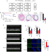
Figure 1
Figure 1
Alliin protects cardiomyocytes against MI injury. (A) Experimental design. The mice were treated with alliin (100 mg/kg) or water 7 days before surgery and for 14 days after surgery. On day 0, surgery was performed, and the mice were sacrificed on day 14. (B) Kaplan-Meier survival curves following MI. (C) Representative Masson's trichrome-stained sections from the sham, MI and alliin-treated MI groups (magnification, x10). (D) Representative echo-cardiography M-mode images from the sham, MI and alliin-treated MI groups. (E) EF and FS percentages in the sham, MI and alliin-treated MI groups. (F) Representative TUNEL staining images (magnification, x40). The data presented in each panel are representative of at least three independent experiments and are presented as the mean ± SEM. *P<0.05, **P<0.01, ***P<0.001. MI, myocardial infarction; EF, ejection fraction; FS, fractional shortening.
Figure 2
Figure 2
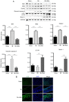
Alliin protects cardiomyocytes against apoptosis and necroptosis. (A) Effect of alliin on the rate of hypoxia-induced apoptosis and necroptosis in H9c2 cells, assessed by flow cytometry. (B) Time course of cell death (Annexin V+ cells). (C) Expression and (D) relative levels of cleaved caspase-3 RIP1, RIP3 and TRAF2 in hypoxia-induced H9c2 cells treated without or with alliin. The data presented in each panel are representative of at least three independent experiments and are presented as the mean ± SEM. *P<0.05, **P<0.01, ***P<0.001. Ctl, control; PI, propidium iodide; RIP, receptor-interacting protein; TRAF2, tumor necrosis factor receptor-associated factor 2.
Figure 3
Figure 3
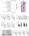
RNA sequencing analysis reveals a role for alliin in autophagy. (A) KEGG pathway analysis of proteins regulated by alliin during hypoxia. (B) Autophagy-related genes regulated by alliin identified by RNA sequencing analysis. (C) Levels of ATG4A, ATG4B, ATG4C, ATG4D, ATG9A and ATG16L2 in primary cardiomyocytes were detected by reverse transcription-quantitative PCR analysis. (D) Expression levels of Beclin 1, LC3, ATG7, p62 and PPARγ in H9c2 cells under hypoxia were analyzed by western blotting, and the (E) effect of alliin was reversed by autophagy inhibitor 3-MA. The data presented in each panel are representative of at least three independent experiments and are presented as the mean ± SEM. *P<0.05, **P<0.01 and ***P<0.001. Ctl, control; LC3, microtubule-associated protein 1 light chain 3; ATG, autophagy-related; PPARγ, peroxisome proliferator-activated receptor γ.
Figure 4
Figure 4
Alliin inhibits hypoxia-induced necroptosis by promoting autophagy. (A) Expression levels of RIP1, RIP3, TRAF2, Beclin 1 and LC3 in the heart following MI were analyzed by western blotting. (B) Representative LC3 staining images (magnification, x40). The data presented in each panel are representative of at least three independent experiments and the data are presented as the mean ± SEM. ***P<0.001. MI, myocardial infarction; RIP, receptor-interacting protein; TRAF2, tumor necrosis factor receptor-associated factor 2; LC3, microtubule-associated protein 1 light chain 3.

Similar articles

Cited by

References

    1. Cahill TJ, Kharbanda RK. Heart failure after myocardial infarction in the era of primary percutaneous coronary intervention: Mechanisms, incidence and identification of patients at risk. World J Cardiol. 2017;9:407–415. doi: 10.4330/wjc.v9.i5.407. - DOI - PMC - PubMed
    1. Zhang T, Zhang Y, Cui M, Jin L, Wang Y, Lv F, Liu Y, Zheng W, Shang H, Zhang J, et al. CaMKII is a RIP3 substrate mediating ischemia- and oxidative stress-induced myocardial necroptosis. Nat Med. 2016;22:175–182. doi: 10.1038/nm.4017. - DOI - PubMed
    1. He S, Huang S, Shen Z. Biomarkers for the detection of necroptosis. Cell Mol Life Sci. 2016;73:2177–2181. doi: 10.1007/s00018-016-2192-3. - DOI - PMC - PubMed
    1. Luedde M, Lutz M, Carter N, Sosna J, Jacoby C, Vucur M, Gautheron J, Roderburg C, Borg N, Reisinger F, et al. RIP3, a kinase promoting necroptotic cell death, mediates adverse remodelling after myocardial infarction. Cardiovasc Res. 2014;103:206–216. doi: 10.1093/cvr/cvu146. - DOI - PubMed
    1. Oerlemans MI, Liu J, Arslan F, den Ouden K, van Middelaar BJ, Doevendans PA, Sluijter JP. Inhibition of RIP1-dependent necrosis prevents adverse cardiac remodeling after myocardial ischemia-reperfusion in vivo. Basic Res Cardiol. 2012;107:270. doi: 10.1007/s00395-012-0270-8. - DOI - PubMed

MeSH terms