. 2019 Dec;44(6):2234-2244.
doi: 10.3892/ijmm.2019.4356. Epub 2019 Sep 27.

Biological effects of melatonin on human adipose‑derived mesenchymal stem cells

Affiliations

Biological effects of melatonin on human adipose‑derived mesenchymal stem cells

June Seok Heo et al. Int J Mol Med. 2019 Dec.

Abstract

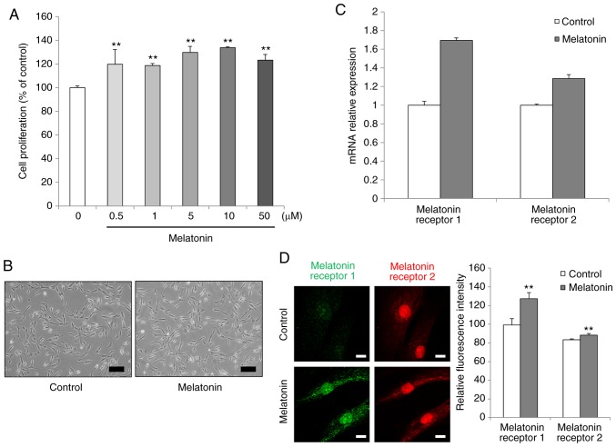
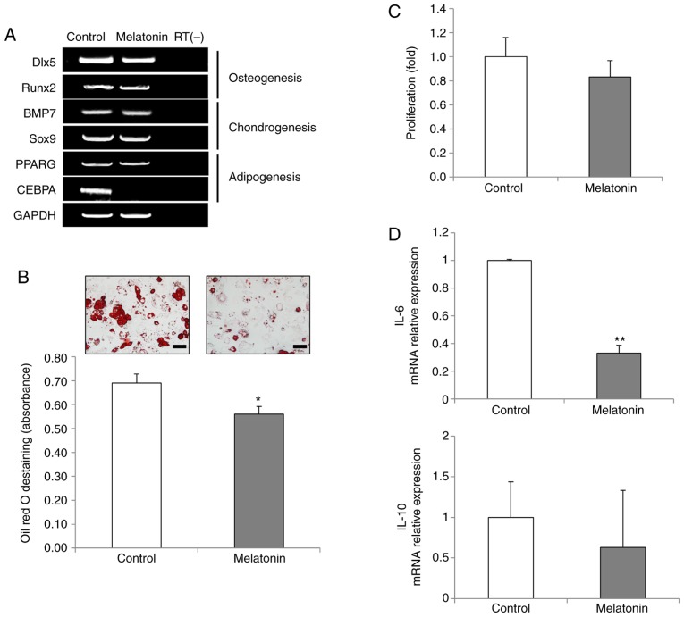
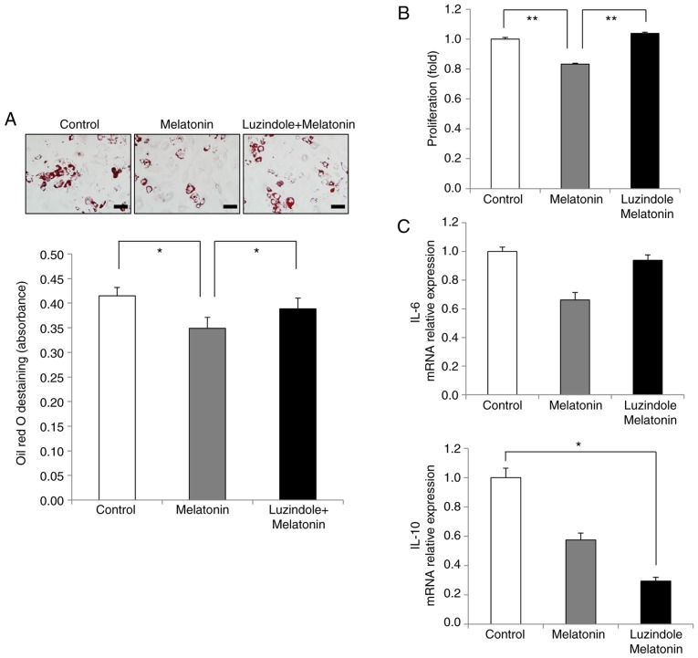
Mesenchymal stem cells (MSCs) are capable of differentiating into other cell types and exhibit immunomodulatory effects. MSCs are affected by several intrinsic and extrinsic signaling modulators, including growth factors, cytokines, extracellular matrix and hormones. Melatonin, produced by the pineal gland, is a hormone that regulates sleep cycles. Recent studies have shown that melatonin improves the therapeutic effects of stem cells. The present study aimed to investigate whether melatonin enhances the biological activities of human adipose‑derived MSCs. The results demonstrated that treatment with melatonin promoted cell proliferation by inducing SRY‑box transcription factor 2 gene expression and preventing replicative senescence. In addition, melatonin exerted anti‑adipogenic effects on MSCs. PCR analysis revealed that the expression of the CCAAT enhancer binding protein a gene, a key transcription factor in adipogenesis, was decreased following melatonin treatment, resulting in reduced adipogenic differentiation in an in vitro assay. The present study also examined the effect of melatonin on the immunomodulatory response using a co‑culture system of human peripheral blood mononuclear cells and MSCs. Activated T cells were strongly inhibited following melatonin exposure compared with those in the control group. Finally, the favorable effects of melatonin on MSCs were confirmed using luzindole, a selective melatonin receptor antagonist. The proliferation‑promoting, anti‑inflammatory effects of melatonin suggested that melatonin‑treated MSCs may be used for effective cell therapy.


Figures

Figure 1
Figure 1
Effects of melatonin on the proliferation of AdMSCs. (A) AdMSCs were cultured with varying concentrations (0.5-50 µM) of melatonin for 3 days. Cell proliferation was determined using an EZ-cytox assay based on WST. (B) Cell morphology images were captured before and after treatment with 10 µM melatonin for 3 days (scale bar, 200 µm; magnification, ×100). (C) Expression of melatonin receptors type 1 and 2 was analyzed by reverse-transcription quantitative PCR. (D) Immunofluorescence staining was performed to examine the protein expression levels of melatonin receptors in untreated cells and melatonin-treated cells (scale bar, 50 µm; magnification, ×63). A representative image of three independent experiments is shown. Data are presented as the mean ± SD of three independent experiments. **P<0.01 compared with control untreated cells. AdMSCs, adipose-derived mesenchymal stem cells.
Figure 2
Figure 2
Effects of melatonin on the senescence of AdMSCs. (A) Gene expression for MSCs and stemness was evaluated through reverse transcription-PCR analysis. RT(-) indicates a negative control using water alone as the reaction input. (B) Self-renewal capacity was assessed through a colony formation assay. (C) Cell senescence was confirmed by β-galactosidase staining after long-term culture (>15 passages). A representative image of three independent experiments is shown (scale bar, 200 µm; magnification, ×100). (D) mRNA expression levels of p21 were analyzed by quantitative PCR. Data are presented as the mean ± SD of three independent experiments. *P<0.05 and **P<0.01. AdMSCs, adipose-derived mesenchymal stem cells; MSCs, mesenchymal stem cells; Oct-4, octamer-binding transcription factor 4; Sox2, SRY-box transcription factor 2; Nanog, nanog homeobox; CFE, colony-forming efficiency.
Figure 3
Figure 3
Effects of melatonin on the differentiation capacity and immunomodulation of AdMSCs. (A) Reverse transcription-PCR analysis of key transcription factors for osteogenesis, chondrogenesis and adipogenesis in untreated cells and in 10 µM melatonin-treated cells. One representative of three independent experiments is shown. RT(-) indicates a negative control using water alone as the reaction input. (B) Adipogenic differentiation capacity was analyzed by Oil Red O staining (scale bar, 50 µm; magnification, ×400). For quantitative analysis, absorbance was measured at 500 nm after destaining. (C) Inhibition of activated mononuclear cells was evaluated through EZ-cytox assays. Phytohaemagglutinin-induced mononuclear cells were co-cultured with untreated or melatonin-treated AdMSCs for 3 days. (D) Relative mRNA expression levels of inflammation-related factors IL-6 and IL-10 were evaluated by quantitative PCR. Data are presented as the mean ± SD of three independent experiments. *P<0.05 and **P<0.01. AdMSCs, adipose-derived mesenchymal stem cells; IL, interleukin; Dlx5, distal-less homeobox 5; Runx2, runt-related transcription factor 2; BMP7, bone morphogenetic protein 7; Sox9, SRY-box transcription factor 9; PPARG, peroxisome proliferator activated receptor γ; CEBPA, CCAAT enhancer binding protein α.
Figure 4
Figure 4
Effects of luzindole on cell proliferation and senescence in melatonin-treated AdMSCs. (A) The expression levels of melatonin receptors type 1 and 2 were determined by quantitative PCR after treatment with 10 µM melatonin and/or 5 µM luzindole for 3 days. (B) The protein expression of melatonin receptors was confirmed using confocal microscopy (scale bar, 50 µm; magnification, ×63). (C) The growth rate of AdMSCs was measured by EZ-Cytox assays after treatment with 10 µM melatonin and/or 5 µM luzindole for 3 days. (D) mRNA expression levels of the p21 gene. (E) Self-renewal capacity was evaluated by a colony formation assay. The number of colonies that formed per 1,000 cells was counted. (F) Senescence-associated β-galactosidase assay of AdMSCs treated with melatonin and/or luzindole after long-term culture. Data are presented as the mean ± SD of three independent experiments. *P<0.05 and **P<0.01, with comparisons indicated by brackets. AdMSCs, adipose-derived mesenchymal stem cells; CFE, colony-forming efficiency.
Figure 5
Figure 5
Effects of luzindole on adipogenesis and immunomodulation in melatonin-treated AdMSCs. (A) Adipogenic differentiation capacity of AdMSCs was evaluated after treatment with 10 µM melatonin and/or 5 µM luzindole for 14 days. One representative of three independent experiments is shown. Absorbance was measured after differentiation for quantitative analysis. (B) Proliferation of mononuclear cells co-cultured with melatonin-treated AdMSCs and melatonin-treated AdMSCs treated with luzindole was determined after 3 days. (C) Relative mRNA expression levels of immunomodulation-related factors IL-6 and IL-10 were analyzed by quantitative PCR. Data are presented as the mean ± SD of three independent experiments. *P<0.05 and **P<0.01, with comparisons indicated by brackets. AdMSCs, adipose-derived mesenchymal stem cells; IL, interleukin.

References

    1. Rivera-Cruz CM, Shearer JJ, Figueiredo Neto M, Figueiredo ML. The immunomodulatory effects of mesenchymal stem cell polarization within the tumor microenvironment niche. Stem Cells Int. 2017;2017:4015039. doi: 10.1155/2017/4015039. - DOI - PMC - PubMed
    1. Si Z, Wang X, Sun C, Kang Y, Xu J, Wang X, Hui Y. Adipose-derived stem cells: Sources, potency, and implications for regenerative therapies. Biomed Pharmacother. 2019;114:108765. doi: 10.1016/j.biopha.2019.108765. - DOI - PubMed
    1. Heo JS, Choi Y, Kim HS, Kim HO. Comparison of molecular profiles of human mesenchymal stem cells derived from bone marrow, umbilical cord blood, placenta and adipose tissue. Int J Mol Med. 2016;37:115–125. doi: 10.3892/ijmm.2015.2413. - DOI - PMC - PubMed
    1. Badimon L, Oñate B, Vilahur G. Adipose-derived mesenchymal stem cells and their reparative potential in ischemic heart disease. Rev Esp Cardiol (Engl Ed) 2015;68:599–611. doi: 10.1016/j.recesp.2015.02.025. - DOI - PubMed
    1. Fernández O, Izquierdo G, Fernández V, Leyva L, Reyes V, Guerrero M, León A, Arnaiz C, Navarro G, Páramo MD, et al. Adipose-derived mesenchymal stem cells (AdMSC) for the treatment of secondary-progressive multiple sclerosis: A triple blinded, placebo controlled, randomized phase I/II safety and feasibility study. PLoS One. 2018;13:e0195891. doi: 10.1371/journal.pone.0195891. - DOI - PMC - PubMed

MeSH terms