

doi: 10.1259/bjr.20190620.
Epub 2019 Oct 1.
Dual-energy CT characterization of winter sports injuries
Affiliations
- PMID: 31573325
- PMCID: PMC7055449
- DOI: 10.1259/bjr.20190620
Item in Clipboard
Dual-energy CT characterization of winter sports injuries
Br J Radiol.
.
Abstract
CT is a readily available imaging modality for cross-sectional characterization of acute musculoskeletal injuries in trauma. Dual-energy CT provides several additional benefits over conventional CT, namely assessment for bone marrow edema, metal artifact reduction, and enhanced assessment of ligamentous injuries. Winter sports such as skiing, snowboarding, and skating can result in high speed and high energy injury mechanisms; dual-energy CT is well suited for the characterization of those injuries.
Figures

A 25-year-old male injured his left hip skiing. Unenhanced DECT image of the hip (1a) showing a non-displaced fracture of the femoral neck (arrow). The 3D bone marrow map (1b) and coronal bone marrow overlay (1c) show corresponding marrow edema (arrowheads), increasing the conspicuity of the fracture. Marrow edema is encoded as green, with normal background marrow blue/purple. The fracture was managed with operative fixation (1d). 3D, three-dimensional; DECT, dual-energy CT.
A 55-year-old male injured his ankle snowboarding. Unenhanced axial and coronal DECT image of the ankle showing a comminuted, non-displaced intra-articular fracture (arrows) of the tibial plafond (2a, 2c). The dual-energy bone marrow overlay shows marrow edema (arrowheads) corresponding with the fractures (2b, 2d). The fracture was managed non-operatively with casting. DECT, dual-energy CT.
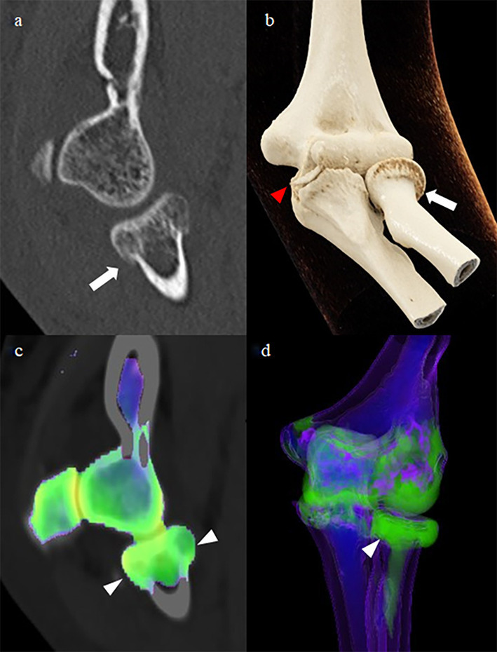
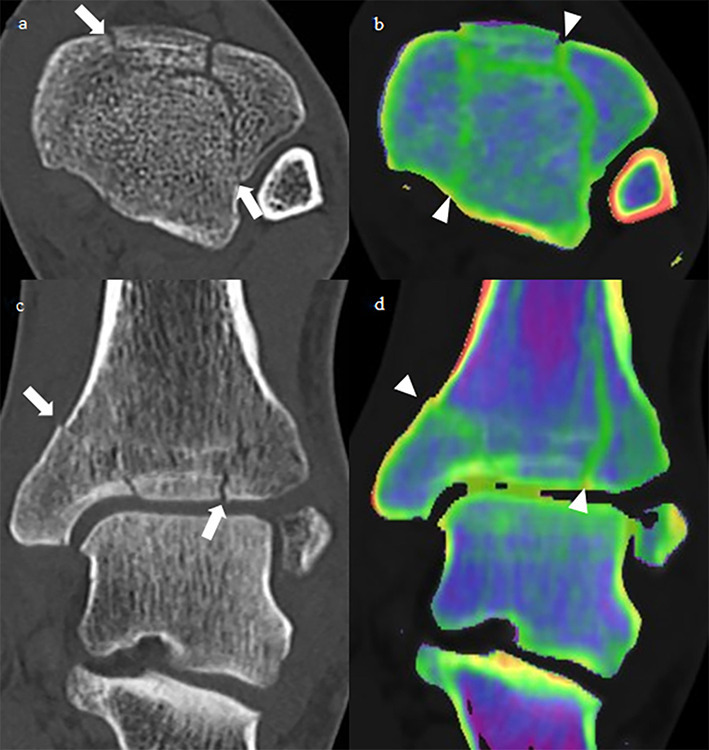
A 53-year-old female injured her elbow skiing. A non-enhanced DECT and 3D cinematic rendering show a radial neck (arrow) and coronoid process (red arrowhead) fractures (3a, 3b). The dual energy marrow overlay and 3D bone marrow map show marrow edema (arrowheads) in the radial head and neck (3c, 3d). There is no apparent edema associated with the coronoid process fracture. Small cortical avulsions often do not show detectable associated edema. 3D, three-dimensional; DECT, dual-energy CT.
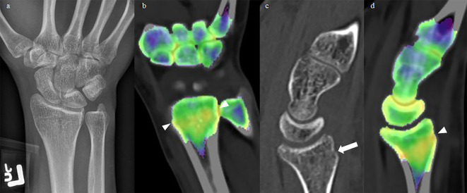
A 28-year-old female injured left wrist snowboarding. The radiograph of the wrist was reported as normal (4a). A DECT was subsequently performed, showing subtle buckling (arrow) of the posterior radial metaphyseal cortex (4c). The bone marrow edema map shows increased water in the radial metaphysis (arrowheads), corresponding with edema in a microtrabecular fracture (4b, 4d). The fracture was managed non-operatively with casting. DECT, dual-energy CT.
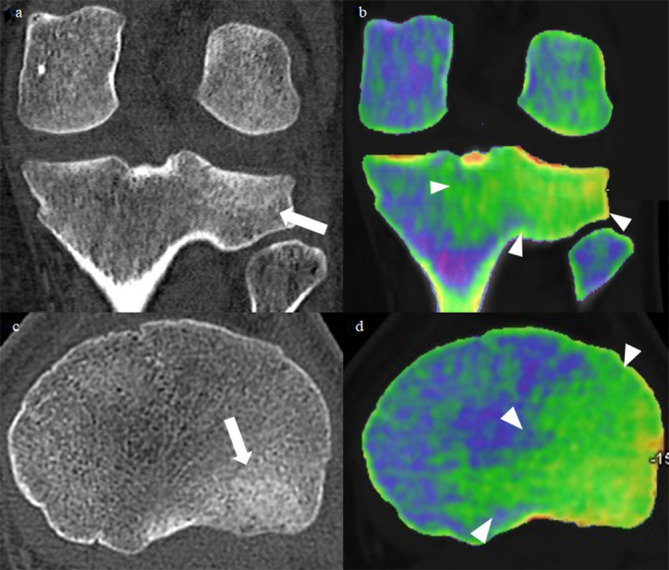
A 23-year-old male injured his knee snowboarding. Unenhanced DECT images of the knee show a subtle fracture (arrows) of the posterolateral tibial plateau (5a, 5c). The bone marrow overlay shows extensive marrow edema (arrowheads) in the posterolateral tibial plateau (5b, 5d). DECT, dual-energy CT.
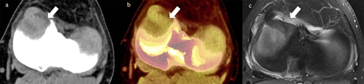
A 36-year-old male felt a “pop” in his knee while ice skating. He presented to the emergency department the next day with a locked, painful knee. Axial unenhanced DECT of the knee at the level of the plateau (6a) shows the medial meniscus flipped into the joint. DECT collagen overlay map (6b) enhances the conspicuity of the flipped meniscus. Axial T2FS MRI image (6c) from the same day confirms the flipped meniscus. Findings were confirmed intra operatively. DECT, dual-energy CT.
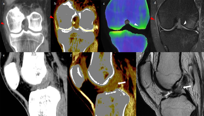
A 24-year-old female injured her knee skiing. Unenhanced coronal DECT (7a) shows medial cruciate ligament (MCL) thickening with periligamentous edema. The tendon mapping application which highlights tightly bundled collagen shows a defect in the proximal MCL (7b, red arrowhead), and indistinctness of the ACL (7f, arrow). The marrow edema overlay (7c) shows focal edema in the lateral femoral condyle (white arrowhead). Coronal T2FS (7d) and sagittal Timages (7g) of the knee show a partial thickness tear of the proximal MCL (red arrowhead), a medial femoral condyle marrow contusion (white arrowhead), and a tear of the ACL (arrow). The patient went on to have an ACL repair.
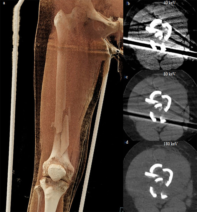
A 53-year-old female injured her right leg snowmobiling. A cinematic rendering of the right femur (8a) shows a comminuted diaphyseal fracture held in an external metal brace. 40, 80 and 180 keV virtual monoenergetic images (8a, b, c) show decreasing beam hardening artifact from the external metallic brace but decreasing soft tissue contrast as the energy level is increased. Her fracture was managed with an intramedullary rod.
Similar articles
-
Self assessment in trauma & orthopaedics II.J R Army Med Corps. 2008 Dec;154(4):247-53. doi: 10.1136/jramc-154-04-09. J R Army Med Corps. 2008. PMID: 19496371 No abstract available.
-
Color-coded virtual non-calcium dual-energy CT for the depiction of bone marrow edema in patients with acute knee trauma: a multireader diagnostic accuracy study.Eur Radiol. 2020 Jan;30(1):141-150. doi: 10.1007/s00330-019-06304-7. Epub 2019 Jul 26. Eur Radiol. 2020. PMID: 31350586
-
Clinical Utility of Dual-Energy CT Analysis of Bone Marrow Edema in Acute Wrist Fractures.AJR Am J Roentgenol. 2018 Apr;210(4):842-847. doi: 10.2214/AJR.17.18673. Epub 2018 Feb 22. AJR Am J Roentgenol. 2018. PMID: 29470155
-
Clinical utility of virtual noncalcium dual-energy CT in imaging of the pelvis and hip.Skeletal Radiol. 2019 Dec;48(12):1833-1842. doi: 10.1007/s00256-019-03243-8. Epub 2019 May 30. Skeletal Radiol. 2019. PMID: 31147733 Review.
-
Essential facets of radiological diagnosis of extremity trauma.CRC Crit Rev Diagn Imaging. 1977 May;9(2):105-205. CRC Crit Rev Diagn Imaging. 1977. PMID: 18319 Review.
Cited by
-
Dual-Energy CT Arthrography: Advanced Muscolo-Skelatal Applications in Clinical Practice.Tomography. 2023 Aug 8;9(4):1471-1484. doi: 10.3390/tomography9040117. Tomography. 2023. PMID: 37624110 Free PMC article. Review.
-
The utility of virtual monochromatic dual-energy computed tomography (DECT) in meniscal imaging: a technical evaluation.Pol J Radiol. 2024 Jul 8;89:e324-e327. doi: 10.5114/pjr/187934. eCollection 2024. Pol J Radiol. 2024. PMID: 39139259 Free PMC article.
-
Reviewing Bone Marrow Edema in Athletes: A Difficult Diagnostic and Clinical Approach.Medicina (Kaunas). 2021 Oct 22;57(11):1143. doi: 10.3390/medicina57111143. Medicina (Kaunas). 2021. PMID: 34833361 Free PMC article. Review.
-
Identification of Non-Traumatic Bone Marrow Oedema: The Pearls and Pitfalls of Dual-Energy CT (DECT).Tomography. 2021 Aug 26;7(3):387-396. doi: 10.3390/tomography7030034. Tomography. 2021. PMID: 34449751 Free PMC article.
-
Imaging of osteoarthritis from the ankle through the midfoot.Skeletal Radiol. 2023 Nov;52(11):2239-2257. doi: 10.1007/s00256-023-04287-7. Epub 2023 Feb 4. Skeletal Radiol. 2023. PMID: 36737484 Free PMC article. Review.
