

Opportunities in Interventional and Diagnostic Imaging by Using High-Performance Low-Field-Strength MRI
- PMID: 31573398
- PMCID: PMC6823617
- DOI: 10.1148/radiol.2019190452
Opportunities in Interventional and Diagnostic Imaging by Using High-Performance Low-Field-Strength MRI
Abstract
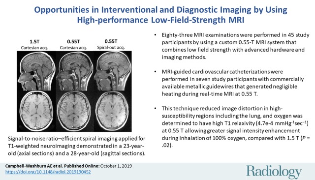
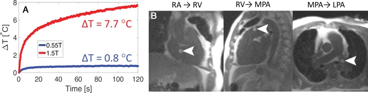
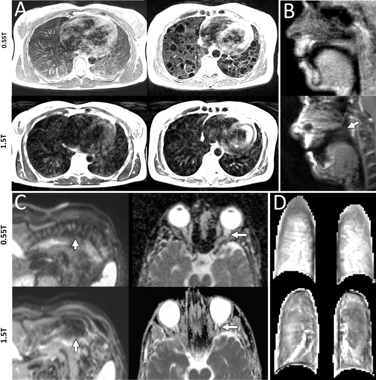
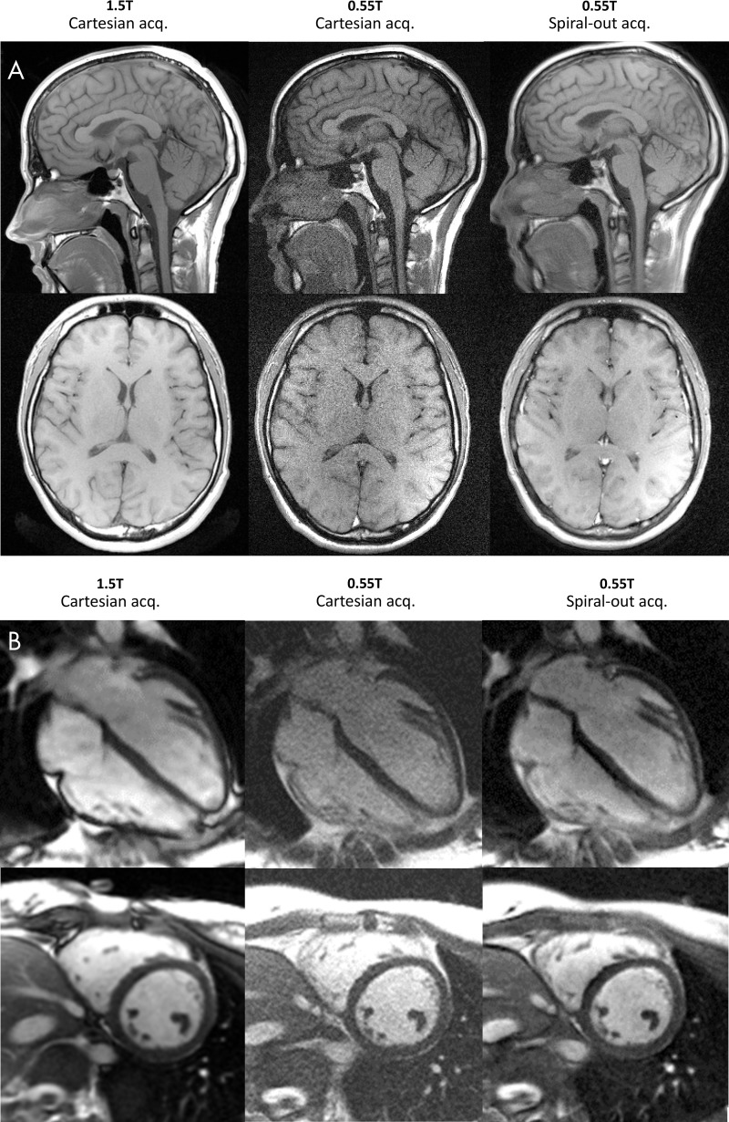
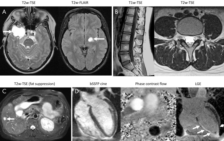
Background Commercial low-field-strength MRI systems are generally not equipped with state-of-the-art MRI hardware, and are not suitable for demanding imaging techniques. An MRI system was developed that combines low field strength (0.55 T) with high-performance imaging technology. Purpose To evaluate applications of a high-performance low-field-strength MRI system, specifically MRI-guided cardiovascular catheterizations with metallic devices, diagnostic imaging in high-susceptibility regions, and efficient image acquisition strategies. Materials and Methods A commercial 1.5-T MRI system was modified to operate at 0.55 T while maintaining high-performance hardware, shielded gradients (45 mT/m; 200 T/m/sec), and advanced imaging methods. MRI was performed between January 2018 and April 2019. T1, T2, and T2* were measured at 0.55 T; relaxivity of exogenous contrast agents was measured; and clinical applications advantageous at low field were evaluated. Results There were 83 0.55-T MRI examinations performed in study participants (45 women; mean age, 34 years ± 13). On average, T1 was 32% shorter, T2 was 26% longer, and T2* was 40% longer at 0.55 T compared with 1.5 T. Nine metallic interventional devices were found to be intrinsically safe at 0.55 T (<1°C heating) and MRI-guided right heart catheterization was performed in seven study participants with commercial metallic guidewires. Compared with 1.5 T, reduced image distortion was shown in lungs, upper airway, cranial sinuses, and intestines because of improved field homogeneity. Oxygen inhalation generated lung signal enhancement of 19% ± 11 (standard deviation) at 0.55 T compared with 7.6% ± 6.3 at 1.5 T (P = .02; five participants) because of the increased T1 relaxivity of oxygen (4.7e-4 mmHg-1sec-1). Efficient spiral image acquisitions were amenable to low field strength and generated increased signal-to-noise ratio compared with Cartesian acquisitions (P < .02). Representative imaging of the brain, spine, abdomen, and heart generated good image quality with this system. Conclusion This initial study suggests that high-performance low-field-strength MRI offers advantages for MRI-guided catheterizations with metal devices, MRI in high-susceptibility regions, and efficient imaging. © RSNA, 2019 Online supplemental material is available for this article. See also the editorial by Grist in this issue.
Figures
Comment in
-
The Next Chapter in MRI: Back to the Future?Radiology. 2019 Nov;293(2):394-395. doi: 10.1148/radiol.2019192011. Epub 2019 Oct 1. Radiology. 2019. PMID: 31577174 No abstract available.
Similar articles
-
Susceptibility artifacts from metallic markers and cardiac catheterization devices on a high-performance 0.55 T MRI system.Magn Reson Imaging. 2021 Apr;77:14-20. doi: 10.1016/j.mri.2020.12.002. Epub 2020 Dec 9. Magn Reson Imaging. 2021. PMID: 33309924 Free PMC article.
-
Right heart catheterization using metallic guidewires and low SAR cardiovascular magnetic resonance fluoroscopy at 1.5 Tesla: first in human experience.J Cardiovasc Magn Reson. 2018 Jun 21;20(1):41. doi: 10.1186/s12968-018-0458-7. J Cardiovasc Magn Reson. 2018. PMID: 29925397 Free PMC article.
-
Segmented nitinol guidewires with stiffness-matched connectors for cardiovascular magnetic resonance catheterization: preserved mechanical performance and freedom from heating.J Cardiovasc Magn Reson. 2015 Nov 30;17:105. doi: 10.1186/s12968-015-0210-5. J Cardiovasc Magn Reson. 2015. PMID: 26620420 Free PMC article.
-
Modern Low-Field MRI of the Musculoskeletal System: Practice Considerations, Opportunities, and Challenges.Invest Radiol. 2023 Jan 1;58(1):76-87. doi: 10.1097/RLI.0000000000000912. Epub 2022 Sep 13. Invest Radiol. 2023. PMID: 36165841 Review.
-
Routine and Advanced Neurologic Imaging at 0.55-T MRI: Opportunities and Challenges.Radiographics. 2025 Mar;45(3):e240076. doi: 10.1148/rg.240076. Radiographics. 2025. PMID: 39946265 Review.
Cited by
-
Novel Techniques in Imaging Congenital Heart Disease: JACC Scientific Statement.J Am Coll Cardiol. 2024 Jan 2;83(1):63-81. doi: 10.1016/j.jacc.2023.10.025. J Am Coll Cardiol. 2024. PMID: 38171712 Free PMC article. Review.
-
Contrast-optimal simultaneous multi-slice bSSFP cine cardiac imaging at 0.55 T.Magn Reson Med. 2023 Feb;89(2):746-755. doi: 10.1002/mrm.29472. Epub 2022 Oct 5. Magn Reson Med. 2023. PMID: 36198043 Free PMC article.
-
[Imaging of the musculoskeletal system using low-field magnetic resonance imaging].Radiologe. 2022 May;62(5):410-417. doi: 10.1007/s00117-022-01000-y. Epub 2022 Apr 13. Radiologe. 2022. PMID: 35416477 Review. German.
-
Evaluation of injuries caused by coronavirus disease 2019 using multi-nuclei magnetic resonance imaging.Magn Reson Lett. 2021 Aug;1(1):2-10. doi: 10.1016/j.mrl.2021.100009. Epub 2021 Aug 8. Magn Reson Lett. 2021. PMID: 35673615 Free PMC article. Review.
-
Non-invasive pressure-volume loops using the elastance model and CMR: a porcine validation at transient pre-loads.Eur Heart J Imaging Methods Pract. 2024 Mar 5;2(1):qyae016. doi: 10.1093/ehjimp/qyae016. eCollection 2024 Jan. Eur Heart J Imaging Methods Pract. 2024. PMID: 38645798 Free PMC article.
References
-
- Konings MK, Bartels LW, Smits HF, Bakker CJ. . Heating around intravascular guidewires by resonating RF waves . J Magn Reson Imaging 2000. ; 12 ( 1 ): 79 – 85 . - PubMed
Publication types
MeSH terms
Substances
Grants and funding
LinkOut - more resources
Full Text Sources
Other Literature Sources
Medical
Miscellaneous
