

Modeling Neuronal Diseases in Zebrafish in the Era of CRISPR
- PMID: 31573887
- PMCID: PMC7324878
- DOI: 10.2174/1570159X17666191001145550
Modeling Neuronal Diseases in Zebrafish in the Era of CRISPR
Abstract
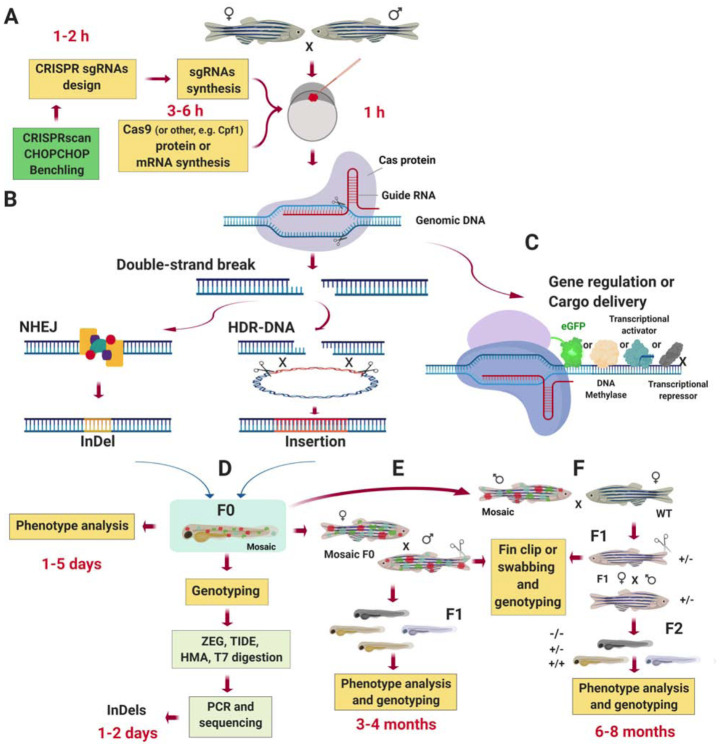
Background: Danio rerio is a powerful experimental model for studies in genetics and development. Recently, CRISPR technology has been applied in this species to mimic various human diseases, including those affecting the nervous system. Zebrafish offer multiple experimental advantages: external embryogenesis, rapid development, transparent embryos, short life cycle, and basic neurobiological processes shared with humans. This animal model, together with the CRISPR system, emerging imaging technologies, and novel behavioral approaches, lay the basis for a prominent future in neuropathology and will undoubtedly accelerate our understanding of brain function and its disorders.
Objective: Gather relevant findings from studies that have used CRISPR technologies in zebrafish to explore basic neuronal function and model human diseases.
Methods: We systematically reviewed the most recent literature about CRISPR technology applications for understanding brain function and neurological disorders in D. rerio. We highlighted the key role of CRISPR in driving forward our understanding of particular topics in neuroscience.
Results: We show specific advances in neurobiology when the CRISPR system has been applied in zebrafish and describe how CRISPR is accelerating our understanding of brain organization.
Conclusion: Today, CRISPR is the preferred method to modify genomes of practically any living organism. Despite the rapid development of CRISPR technologies to generate disease models in zebrafish, more efforts are needed to efficiently combine different disciplines to find the etiology and treatments for many brain diseases.
Keywords: Brain disease models; CRISPR; Danio rerio; genome engineering; optogenetics; zebrafish..
Copyright© Bentham Science Publishers; For any queries, please email at epub@benthamscience.net.
Figures


Similar articles
-
Understanding neurobehavioral genetics of zebrafish.J Neurogenet. 2020 Mar-Jun;34(2):203-215. doi: 10.1080/01677063.2019.1698565. Epub 2020 Jan 5. J Neurogenet. 2020. PMID: 31902276 Review.
-
Effective CRISPR/Cas9-based nucleotide editing in zebrafish to model human genetic cardiovascular disorders.Dis Model Mech. 2018 Oct 18;11(10):dmm035469. doi: 10.1242/dmm.035469. Dis Model Mech. 2018. PMID: 30355756 Free PMC article.
-
Efficient Production and Identification of CRISPR/Cas9-generated Gene Knockouts in the Model System Danio rerio.J Vis Exp. 2018 Aug 28;(138):56969. doi: 10.3791/56969. J Vis Exp. 2018. PMID: 30222157 Free PMC article.
-
CRISPR/Cas9 in zebrafish: an efficient combination for human genetic diseases modeling.Hum Genet. 2017 Jan;136(1):1-12. doi: 10.1007/s00439-016-1739-6. Epub 2016 Nov 2. Hum Genet. 2017. PMID: 27807677 Free PMC article. Review.
-
Multiple genome modifications by the CRISPR/Cas9 system in zebrafish.Genes Cells. 2014 Jul;19(7):555-64. doi: 10.1111/gtc.12154. Epub 2014 May 22. Genes Cells. 2014. PMID: 24848337
Cited by
-
Zebrafish as a tool for autism research: unraveling the roles of Shank3, Cntnap2, Neuroligin3, and Arid1b in synaptic and behavioral abnormalities.Neurogenetics. 2025 Jun 6;26(1):48. doi: 10.1007/s10048-025-00828-5. Neurogenetics. 2025. PMID: 40478461 Review.
-
Mutants of the Zebrafish K+ Channel Hcn2b Exhibit Epileptic-like Behaviors.Int J Mol Sci. 2021 Oct 25;22(21):11471. doi: 10.3390/ijms222111471. Int J Mol Sci. 2021. PMID: 34768904 Free PMC article.
-
Potential contributions of the intrinsic retinal oscillations recording using non-invasive electroretinogram to bioelectronics.Front Cell Neurosci. 2024 Jan 8;17:1224558. doi: 10.3389/fncel.2023.1224558. eCollection 2023. Front Cell Neurosci. 2024. PMID: 38269118 Free PMC article.
-
PHLPP1 promotes neutral lipid accumulation through AMPK/ChREBP-dependent lipid uptake and fatty acid synthesis pathways.iScience. 2022 Jan 12;25(2):103766. doi: 10.1016/j.isci.2022.103766. eCollection 2022 Feb 18. iScience. 2022. PMID: 35141506 Free PMC article.
-
Zebrafish as a Tool in the Study of Sleep and Memory-related Disorders.Curr Neuropharmacol. 2022 Mar 4;20(3):540-549. doi: 10.2174/1570159X19666210712141041. Curr Neuropharmacol. 2022. PMID: 34254919 Free PMC article. Review.
References
Publication types
MeSH terms
LinkOut - more resources
Full Text Sources
Medical
