. 2020 Jan:28:101328.
doi: 10.1016/j.redox.2019.101328. Epub 2019 Sep 20.

Insight into the mechanism of ferroptosis inhibition by ferrostatin-1

Affiliations

Insight into the mechanism of ferroptosis inhibition by ferrostatin-1

Giovanni Miotto et al. Redox Biol. 2020 Jan.

Abstract

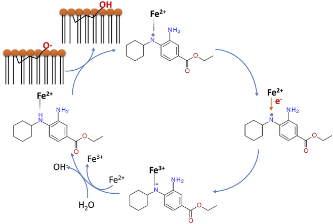
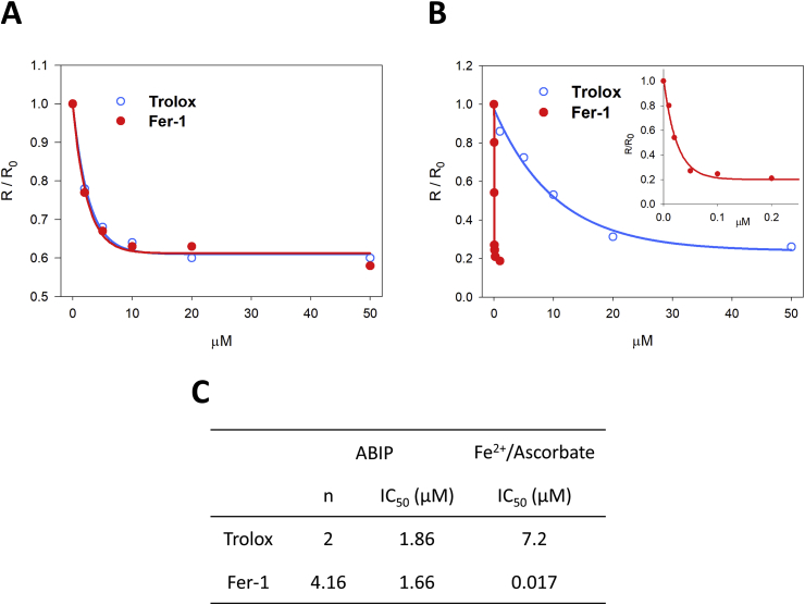
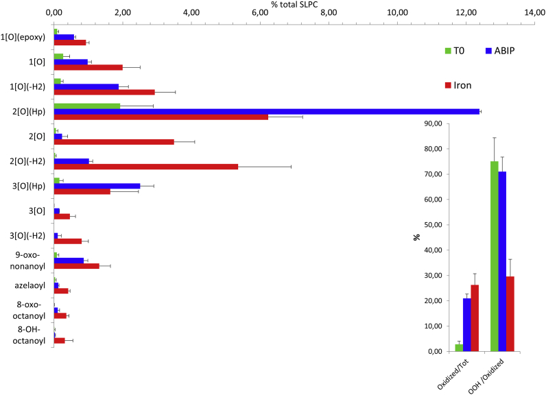
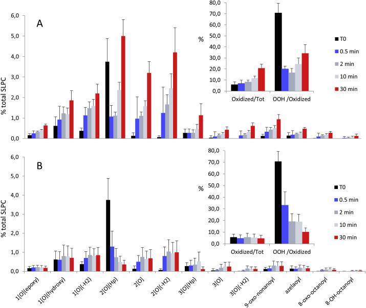
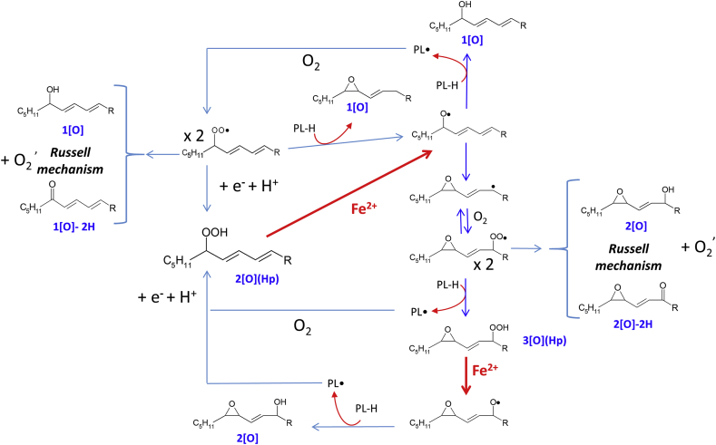
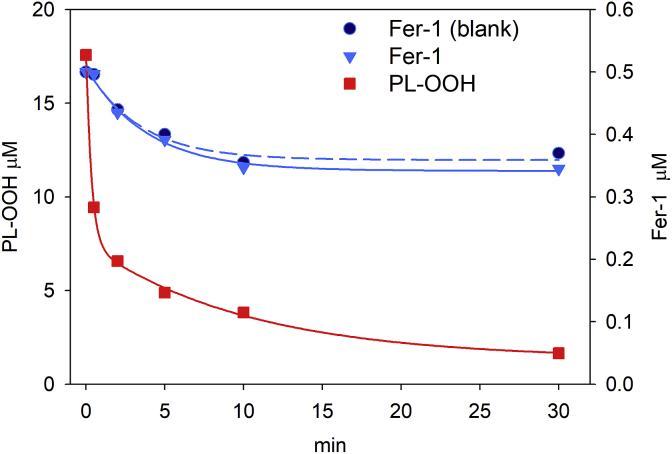
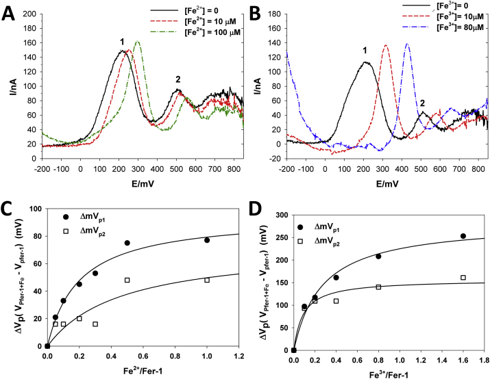
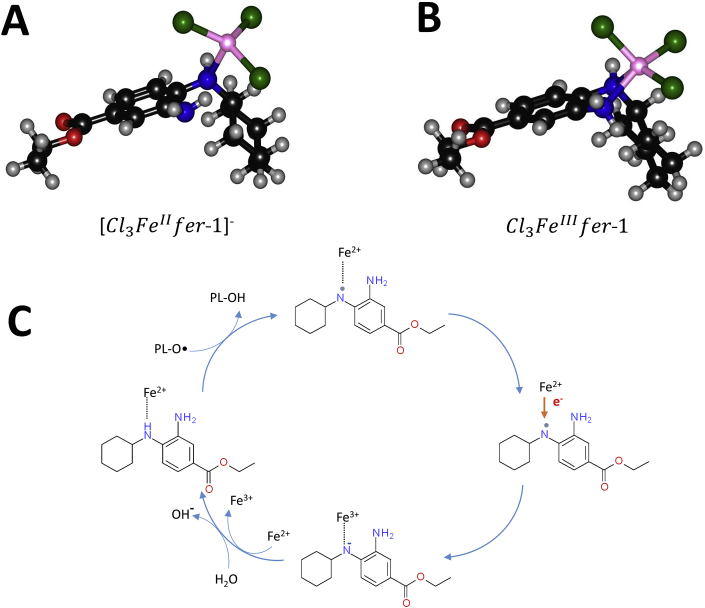
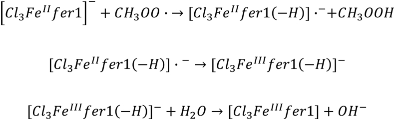
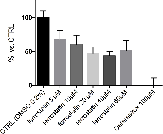
Ferroptosis is a form of cell death primed by iron and lipid hydroperoxides and prevented by GPx4. Ferrostatin-1 (fer-1) inhibits ferroptosis much more efficiently than phenolic antioxidants. Previous studies on the antioxidant efficiency of fer-1 adopted kinetic tests where a diazo compound generates the hydroperoxyl radical scavenged by the antioxidant. However, this reaction, accounting for a chain breaking effect, is only minimally useful for the description of the inhibition of ferrous iron and lipid hydroperoxide dependent peroxidation. Scavenging lipid hydroperoxyl radicals, indeed, generates lipid hydroperoxides from which ferrous iron initiates a new peroxidative chain reaction. We show that when fer-1 inhibits peroxidation, initiated by iron and traces of lipid hydroperoxides in liposomes, the pattern of oxidized species produced from traces of pre-existing hydroperoxides is practically identical to that observed following exhaustive peroxidation in the absence of the antioxidant. This supported the notion that the anti-ferroptotic activity of fer-1 is actually due to the scavenging of initiating alkoxyl radicals produced, together with other rearrangement products, by ferrous iron from lipid hydroperoxides. Notably, fer-1 is not consumed while inhibiting iron dependent lipid peroxidation. The emerging concept is that it is ferrous iron itself that reduces fer-1 radical. This was supported by electroanalytical evidence that fer-1 forms a complex with iron and further confirmed in cells by fluorescence of calcein, indicating a decrease of labile iron in the presence of fer-1. The notion of such as pseudo-catalytic cycle of the ferrostatin-iron complex was also investigated by means of quantum mechanics calculations, which confirmed the reduction of an alkoxyl radical model by fer-1 and the reduction of fer-1 radical by ferrous iron. In summary, GPx4 and fer-1 in the presence of ferrous iron, produces, by distinct mechanism, the most relevant anti-ferroptotic effect, i.e the disappearance of initiating lipid hydroperoxides.

Keywords: Alkoxyl radical; Antioxidant; Ferroptosis; Ferrostatin-1; GPx4; Lipid peroxidation.


Figures

Image 1
Graphical abstract
Fig. 1
Fig. 1
Inhibition of lipid peroxidation induced with iron or diazo in liposome. The dependence of initial oxygen consumption rate ratio R/R0 from trolox (blue open circle) and fer-1 (red full circle, also expanded into the inset) concentration was fitted to Eq (1) (See Materials and Methods). Liposome solution containing 0.1 mM phospholipids (SLPC:SLPE/1:1), different amount of trolox or fer-1 was oxidized by 8 mM ABIP (panel A) or 30 μM ascorbic acid and 4 μM FeSO4 (panel B) in 0.1 M Tris, 0.15 M KCl at pH 7.0, at 37 °C. In (C) IC50 values calculated from panels A and B are reported. (For interpretation of the references to colour in this figure legend, the reader is referred to the Web version of this article.)
Fig. 2
Fig. 2
Lipid oxidation pattern induced by diazo compounds or Fe2+/ascorbate. Oxidized lipids pattern, relative to 1-stearoyl-2-linoleoyl-sn-glycero-3-phosphocoline (SLPC), of a fresh liposome suspension containing 0.1 mM phospholipids (SLPC:SLPE/1:1), was measured at time 0 (T0) and after 30 min of incubation with ABIP 8 mM or Fe2+ 4 μM plus Ascorbate 30 μM. In the inset are summarized the percentage of the overall oxidized species versus total amount of SPLC (left) and the percentage of lipo-hydroperoxides versus the sum of oxidized species (right). Values represent the mean ± standard deviation of 20 independent experiments.
Fig. 3
Fig. 3
Time course of Fe2+ induced SLPC oxidation pattern, and fer-1 effect. Liposomes suspension, containing 0.1 mM phospholipids (SLPC:SLPE/1:1), was incubated for 30 min with Fe2+ 4 μM and Ascorbate 30 μM in absence (A) or in presence (B) of 0.5 μM fer-1. Samples were withdrawn before additions (T0) and at 0.5, 2, 10, 30 min after additions. In the insets the percentage of the overall oxidized species versus total amount of SPLC (left) and the percentage of lipo-hydroperoxides versus the sum of oxidized species (right) are summarized. Values represent the mean ± standard deviation of 3 independent experiments.
Fig. 4
Fig. 4
Schematic outline of Fe2+ induced lipid peroxidation mechanism.
Fig. 5
Fig. 5
Lipid hydroperoxide decomposition and fer-1 consumption in the presence of iron. A solution containing 0.1 mM phospholipids (SLPC:SLPE/1:1) in 0.1 M Tris, 0.15 M KCl at pH 7.4, enriched with hydroperoxides by a pretreatment with Alox15 for 60min, was treated with 0.5 μM fer-1, 30 μM ascorbic acid and 4 μM FeSO4, at 37 °C. Time course of total PL-OOH (square ■) and fer-1 (triangle▼) were quantified by Mass Spectrometry. Blank was fer-1 in buffer (circle ●).
Fig. 6
Fig. 6
Effect of ferric and ferrous ions on the anodic peak potential values of fer-1. Differential pulse voltammograms of fer-1 in the absence and presence of different ferrous (A) and ferric (B) ion concentrations show the anodic shift of peak potential values of fer-1 induced by iron. Plots of the shifts of the peak potentials (ΔVp = VpFer-1+Fe – VpFer-1) on the Fe2+/fer-1 (C) and Fe3+/fer-1 (D) are shown; continuous lines are the results of the best fitting of the hyperbola equation for the experimental data, by non-linear regression analysis.
Fig. 7
Fig. 7
Molecular structures of iron complexes with ferrostatin-1 fully optimized at M06-L/6–31G(d,p), LanL2DZ level of theory. Fer-1/FeII (A) and fer-1/FeIII (B) complexes are shown in (A) and (B) respectively. In (C) a schematic view of fer-1 scavenging the lipid alkoxy radicals (PL-O·) and reduction of fer-1 radical by ferrous iron is depicted.
Scheme 1
Scheme 1
Elementary steps of radical reduction via HAT from fer-1.
Fig. 8
Fig. 8
Fer-1 binds iron of Labile Iron Pool (LIP). Relative LIP was expressed as calcein Mean Fluorescence Intensity (MFI) in treated cells vs. control. Cells incubated with Deferasirox 100 μM served as baseline. The values are the mean ± standard deviation (SD) of 2 experiment in triplicate.

References

    1. Dixon S.J., Lemberg K.M., Lamprecht M.R., Skouta R., Zaitsev E.M., Gleason C.E. Ferroptosis: an iron-dependent form of nonapoptotic cell death. Cell. 2012;149:1060–1072. - PMC - PubMed
    1. Guiney S.J., Adlard P.A., Bush A.I., Finkelstein D.I., Ayton S. Ferroptosis and cell death mechanisms in Parkinson's disease. Neurochem. Int. 2017;104:34–48. - PubMed
    1. Conrad M., Angeli J.P., Vandenabeele P., Stockwell B.R. Regulated necrosis: disease relevance and therapeutic opportunities. Nat. Rev. Drug Discov. 2016;15:348–366. - PMC - PubMed
    1. Magtanong L., Dixon S.J. Ferroptosis and brain injury. Dev. Neurosci. 2019:1–14. - PMC - PubMed
    1. Zhu H., Santo A., Jia Z., Robert Li Y. GPx4 in Bacterial Infection and Polymicrobial Sepsis: Involvement of Ferroptosis and Pyroptosis. React. Oxyg. Species (Apex) 2019;7:154–160. - PMC - PubMed

Publication types

MeSH terms

LinkOut - more resources