

Management of giant benign fibrous histiocytoma in the spinal region with pleural involvement: A case report
- PMID: 31574815
- PMCID: PMC6775359
- DOI: 10.1097/MD.0000000000017144
Management of giant benign fibrous histiocytoma in the spinal region with pleural involvement: A case report
Abstract
Rationale: Benign fibrous histiocytoma with pleural involvement in spinal region is a highly unusual disease with no standard curative managements yet. The objective of this study is to report an extremely rare case of a giant benign fibrous histiocytoma with pleural involvement in spinal region successfully operated by posterior spinal surgery. The management of these unique cases has yet to be well-documented.
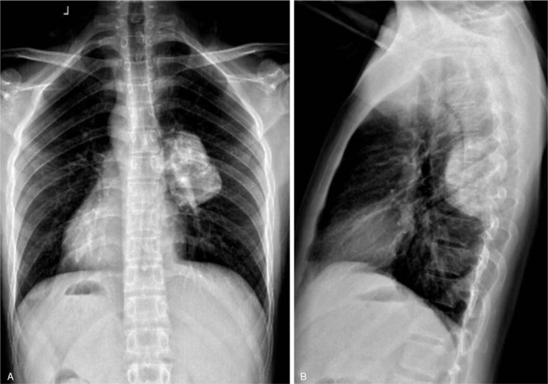
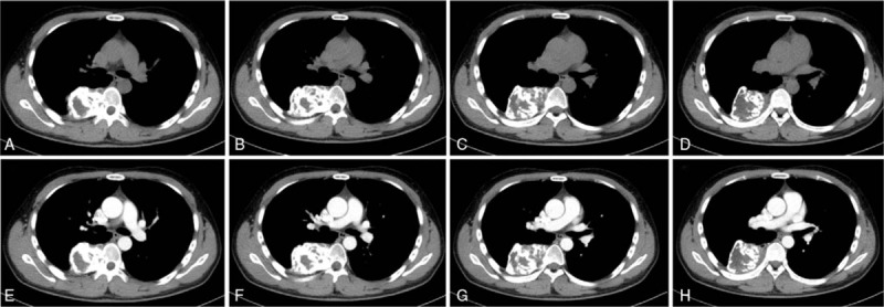
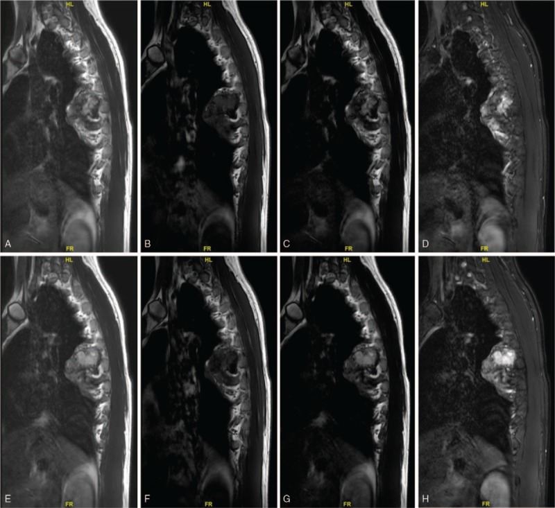
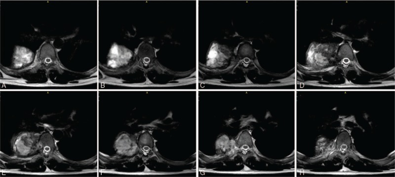
Patient concerns: A 23-year-old man presented with a 2-month history of continuous and progressive back pain. A giant, expanding lesion of the T7 vertebral and paraspinal region with pleural involvement was identified.
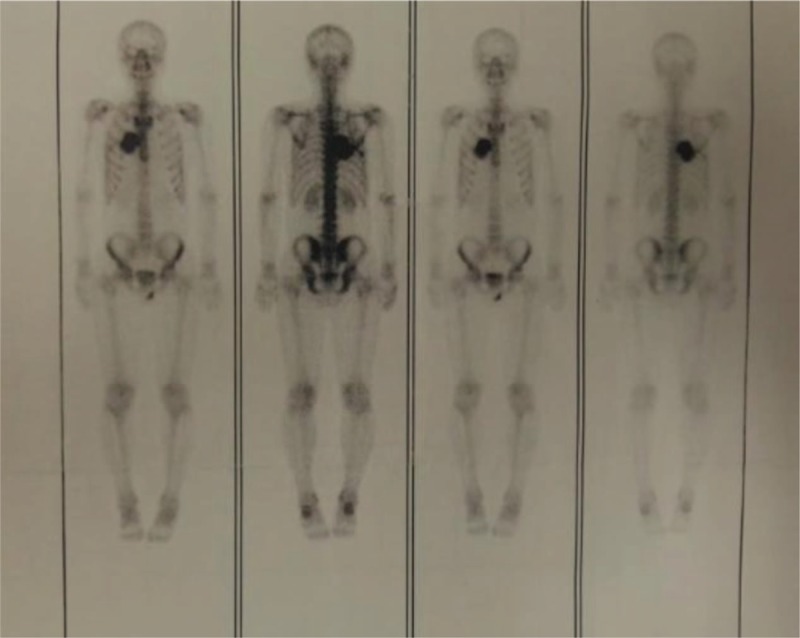
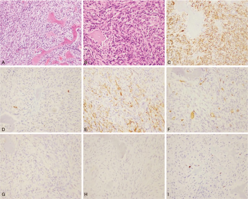
Diagnoses: Computed tomography scan and magnetic resonance imaging of spine showed expanding lesion of the T7 vertebral and paraspinal region involving the right thoracic cavity, which presented as a solid tumor. Postoperative pathology confirmed the diagnosis of thoracic benign fibrous histiocytoma.
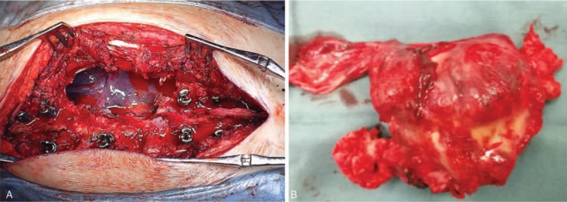
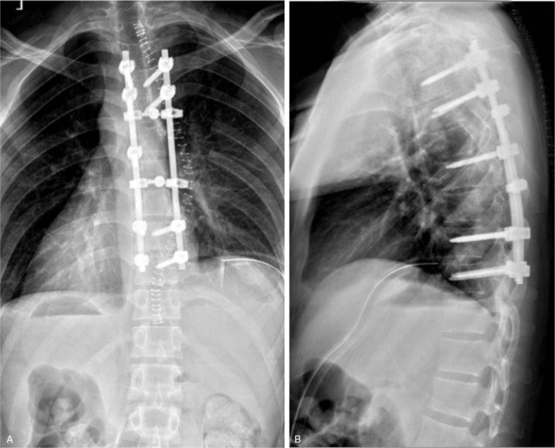
Interventions: The patient underwent thoracic spinal canal decompression, complete tumor resection, pleural defect repair, and T4 to T10 internal fixation procedure via a posterior approach.
Outcomes: The patient's symptom improved significantly after the surgery, and the postoperative period was uneventful at the 2-year follow-up visit. There were no complications associated with the operation during the follow-up period.
Lessons: In summary, the tumor's clinical features, imaging results, and pathological characteristics are unique. Combined efforts of specialists from orthopedics, thoracic surgery, neurosurgery, pathology, and medical oncology led to the successful diagnosis and management of this patient. Giant benign fibrous histiocytoma with pleural involvement in spinal region, although rare, should be part of the differential diagnosis when the patient presents with back pain and radiculopathy. We recommend the posterior or 1-stage anterior-posterior combined approach for complete resection of the giant thoracic benign fibrous histiocytoma when the tumor has caused severe symptoms or neurological deficits.
Conflict of interest statement
The authors have no conflicts of interest to disclose.
Figures
Similar articles
-
Surgical treatment of giant chordoma in the thoracic spine combining thoracoscopic and posterior spinal surgery: A case report.Medicine (Baltimore). 2019 Aug;98(35):e16990. doi: 10.1097/MD.0000000000016990. Medicine (Baltimore). 2019. PMID: 31464948 Free PMC article.
-
Surgical treatment of malignant paraganglioma with spinal invasion in a juvenile patient: A case report.Medicine (Baltimore). 2019 Sep;98(39):e17145. doi: 10.1097/MD.0000000000017145. Medicine (Baltimore). 2019. PMID: 31574816 Free PMC article.
-
Surgical management of spinal metastases of thymic carcinoma: A case report and literature review.Medicine (Baltimore). 2019 Jan;98(3):e14198. doi: 10.1097/MD.0000000000014198. Medicine (Baltimore). 2019. PMID: 30653174 Free PMC article. Review.
-
Surgical treatment of chondrosarcoma of the sacrum with cement augmentation: A case report.Medicine (Baltimore). 2019 Dec;98(50):e18413. doi: 10.1097/MD.0000000000018413. Medicine (Baltimore). 2019. PMID: 31852164 Free PMC article.
-
Benign fibrous histiocytoma of the posterior arch of C1 in a 6-year-old boy: a case report.Spine (Phila Pa 1976). 2003 Sep 15;28(18):E359-63. doi: 10.1097/01.BRS.0000091337.93304.FA. Spine (Phila Pa 1976). 2003. PMID: 14501936 Review.
Cited by
-
Primary extradural tumors of the spinal column: A comprehensive treatment guide for the spine surgeon based on the 5th Edition of the World Health Organization bone and soft-tissue tumor classification.J Craniovertebr Junction Spine. 2021 Oct-Dec;12(4):336-360. doi: 10.4103/jcvjs.jcvjs_115_21. Epub 2021 Dec 11. J Craniovertebr Junction Spine. 2021. PMID: 35068816 Free PMC article. Review.
References
-
- Skunda R, Puckett T, Martin M, et al. 14-year-old boy with mild antecedent neck pain in setting of acute trauma: a rare case of benign fibrous histiocytoma of the spine. Am J Orthop (Belle Mead NJ) 2016;45:E148–52. - PubMed
-
- Donati F, Proietti L, Burrofato A, et al. Intraspinal extradural benign fibrous histiocytoma of the lumbar spine in a pediatric patient. Case report and literature review. Childs Nerv Syst 2016;32:1549–53. - PubMed
-
- Khor YM, Yan X. Benign fibrous histiocytoma of the thoracic spine as the cause of pyrexia of unknown origin identified by positron emission tomography/computed tomography. Spine J 2015;15:1691–2. - PubMed
-
- Yang M, Wang XB, Li J, et al. Surgical treatment of large abdominally involved primary dumbbell tumor in the lumbar region. J Spinal Disord Tech 2014;27:E268–75. - PubMed
Publication types
MeSH terms
LinkOut - more resources
Full Text Sources
Research Materials
