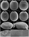
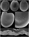
Morphology and Phylogenetics of Benthic Prorocentrum Species (Dinophyceae) from Tropical Northwestern Australia
- PMID: 31574958
- PMCID: PMC6833055
- DOI: 10.3390/toxins11100571
Morphology and Phylogenetics of Benthic Prorocentrum Species (Dinophyceae) from Tropical Northwestern Australia
Abstract
Approximately 70 species of Prorocentrum are known, of which around 30 species are associated with benthic habitats. Some produce okadaic acid (OA), dinophysistoxin (DTX) and their derivatives, which are involved in diarrhetic shellfish poisoning. In this study, we isolated and characterized Prorocentrum concavum and P. malayense from Broome in north Western Australia using light and scanning electron microscopy as well as molecular sequences of large subunit regions of ribosomal DNA, marking the first record of these species from Australian waters. The morphology of the motile cells of P. malayense was similar to P. concavum in the light microscopy, but differed by the smooth thecal surface, the pore pattern and the production of mucous stalk-like structures and a hyaline sheath around the non-motile cells. P. malayense could also be differentiated from other closely related species, P. leve and P. foraminosum, despite the similarity in thecal surface and pore pattern, by its platelet formula and morphologies. We tested the production of OA and DTXs from both species, but found that they did not produce detectable levels of these toxins in the given culturing conditions. This study aids in establishing more effective monitoring of potential harmful algal taxa in Australian waters for aquaculture and recreational purposes.
Keywords: Prorocentrum; benthic dinoflagellates; phylogeny; taxonomy.
Conflict of interest statement
The authors declare no conflicts of interest.
Figures


Similar articles
-
Taxonomy and toxicity of Prorocentrum from Perhentian Islands (Malaysia), with a description of a non-toxigenic species Prorocentrum malayense sp. nov. (Dinophyceae).Harmful Algae. 2019 Mar;83:95-108. doi: 10.1016/j.hal.2019.01.007. Epub 2019 Mar 15. Harmful Algae. 2019. PMID: 31097256
-
Effects of nitrogen concentration and cold temperature on DSP-toxin concentrations in the dinoflagellate Prorocentrum lima (Prorocentrales, Dinophyceae).Nat Toxins. 1994;2(5):263-70. doi: 10.1002/nt.2620020504. Nat Toxins. 1994. PMID: 7866661
-
Morphology and phylogeny of Prorocentrum caipirignum sp. nov. (Dinophyceae), a new tropical toxic benthic dinoflagellate.Harmful Algae. 2017 Dec;70:73-89. doi: 10.1016/j.hal.2017.11.001. Epub 2017 Nov 10. Harmful Algae. 2017. PMID: 29169570
-
The Mechanism of Diarrhetic Shellfish Poisoning Toxin Production in Prorocentrum spp.: Physiological and Molecular Perspectives.Toxins (Basel). 2016 Sep 22;8(10):272. doi: 10.3390/toxins8100272. Toxins (Basel). 2016. PMID: 27669302 Free PMC article. Review.
-
Taxonomy and abundance of epibenthic Prorocentrum (Dinophyceae) species from the tropical and subtropical Southwest Atlantic Ocean including a review of their global diversity and distribution.Harmful Algae. 2023 Aug;127:102470. doi: 10.1016/j.hal.2023.102470. Epub 2023 Jun 10. Harmful Algae. 2023. PMID: 37544670 Review.
Cited by
-
Effect of Different Species of Prorocentrum Genus on the Japanese Oyster Crassostrea gigas Proteomic Profile.Toxins (Basel). 2021 Jul 20;13(7):504. doi: 10.3390/toxins13070504. Toxins (Basel). 2021. PMID: 34357976 Free PMC article.
-
Ribosomal DNA Sequence-Based Taxonomy and Antimicrobial Activity of Prorocentrum spp. (Dinophyceae) from Mauritius Coastal Waters, South-West Indian Ocean.Mar Drugs. 2023 Mar 28;21(4):216. doi: 10.3390/md21040216. Mar Drugs. 2023. PMID: 37103354 Free PMC article.
-
Mucus-Trap-Assisted Feeding Is a Common Strategy of the Small Mixoplanktonic Prorocentrum pervagatum and P. cordatum (Prorocentrales, Dinophyceae).Microorganisms. 2023 Jul 1;11(7):1730. doi: 10.3390/microorganisms11071730. Microorganisms. 2023. PMID: 37512902 Free PMC article.
References
-
- Massana R., Gobet A., Audic S., Bass D., Bittner L., Boutte C., Chambouvet A., Christen R., Claverie J.M., Decelle J. Marine protist diversity in European coastal waters and sediments as revealed by high throughput sequencing. Environ. Microbiol. 2015;17:4035–4049. doi: 10.1111/1462-2920.12955. - DOI - PubMed
-
- Murray S. Diversity and Phylogenetics of Sand-Dwelling Dino-Flagellates. VDM Verlag Dr. Müller Saarbrücken; Riga, Latvia: 2009.
-
- Hoppenrath M., Murray S.A., Chomérat N., Horiguchi T. Marine Benthic Dinoflagellates-Unveiling Their Worldwide Biodiversity. Schweizerbart/Borntraeger; Stuttgart, Germany: 2014.
Publication types
MeSH terms
Substances
LinkOut - more resources
Full Text Sources

