. 2019 Sep 30;11(10):1601.
doi: 10.3390/polym11101601.

3D Bio-Printing of CS/Gel/HA/Gr Hybrid Osteochondral Scaffolds

Affiliations

3D Bio-Printing of CS/Gel/HA/Gr Hybrid Osteochondral Scaffolds

Xueyan Hu et al. Polymers (Basel). .

Abstract

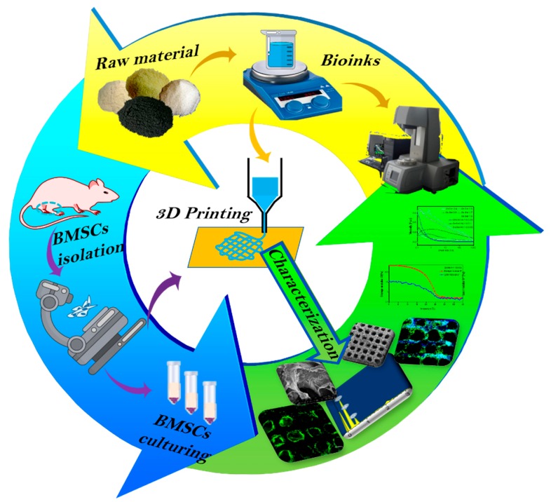
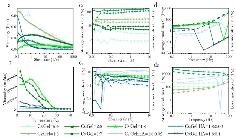
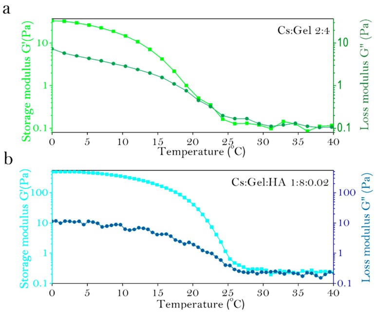
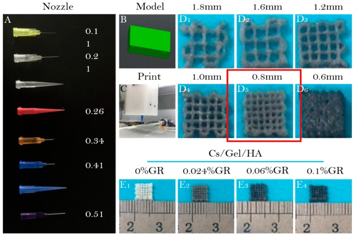
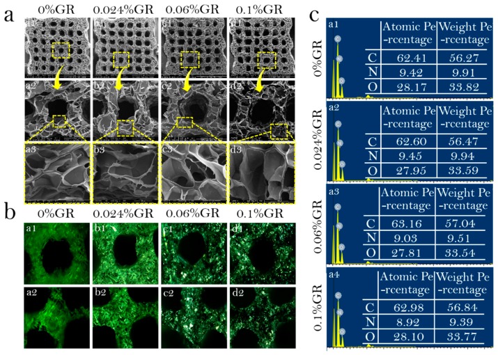
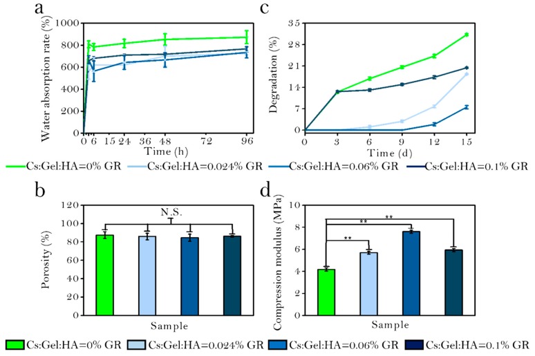
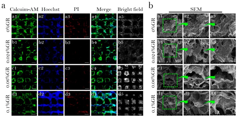
Cartilage is an important tissue contributing to the structure and function of support and protection in the human body. There are many challenges for tissue cartilage repair. However, 3D bio-printing of osteochondral scaffolds provides a promising solution. This study involved preparing bio-inks with different proportions of chitosan (Cs), Gelatin (Gel), and Hyaluronic acid (HA). The rheological properties of each bio-ink was used to identify the optimal bio-ink for printing. To improve the mechanical properties of the bio-scaffold, Graphene (GR) with a mass ratio of 0.024, 0.06, and 0.1% was doped in the bio-ink. Bio-scaffolds were prepared using 3D printing technology. The mechanical strength, water absorption rate, porosity, and degradation rate of the bio-scaffolds were compared to select the most suitable scaffold to support the proliferation and differentiation of cells. P3 Bone mesenchymal stem cells (BMSCs) were inoculated onto the bio-scaffolds to study the biocompatibility of the scaffolds. The results of SEM showed that the Cs/Gel/HA scaffolds with a GR content of 0, 0.024, 0.06, and 0.1% had a good three-dimensional porous structure and interpenetrating pores, and a porosity of more than 80%. GR was evenly distributed on the scaffold as observed by energy spectrum analyzer and polarizing microscope. With increasing GR content, the mechanical strength of the scaffold was enhanced, and pore walls became thicker and smoother. BMSCs were inoculated on the different scaffolds. The cells distributed and extended well on Cs/Gel/HA/GR scaffolds. Compared to traditional methods in tissue-engineering, this technique displays important advantages in simulating natural cartilage with the ability to finely control the mechanical and chemical properties of the scaffold to support cell distribution and proliferation for tissue repair.

Keywords: 3D printing; bio-ink; cartilage repair; chitosan/gelatin/hyaluronic acid; graphene.


Conflict of interest statement

The authors have no conflict of interest.

Figures

Figure 1
Figure 1
Schematic illustration of the general study design. Yellow area: Preparation of Cs/Gel/HA/GR bio-inks and production of scaffolds using 3D bio-printing. Green area: Characterizations of Cs/Gel/HA/GR cartilage scaffolds using scanning electron microscope (SEM) and live/dead staining of cells.
Figure 2
Figure 2
Rheological properties of bio-ink. (a): Shear rate, (b): Viscosity profile, (c1,c2): Strain, and (d1,d2): Frequency.
Figure 3
Figure 3
Gel point for various preparations of Cs:Gel:HA bio-ink, (a): Cs:Gel = 2:4, (b): Cs:Gel:HA = 1:8:0.02.
Figure 4
Figure 4
3D bio-printing using bio-ink. (A): Needles with varying diameters; (B): 3D printing model of bio-scaffold; (C): Images of the 3D printer; (D1D6): Appearance of bio-scaffolds with different filling distances at (D1) 1.8 mm, (D2) 1.6 mm, (D3) 1.2 mm, (D4) 1.0 mm, (D5) 0.8 mm, and (D6) 0.6 mm; Appearance of (E1) Cs/Gel/HA composite scaffold, (E2) Cs/Gel/HA/0.024%GR composite scaffold, (E3) Cs/Gel/HA/0.06%GR composite scaffold, and (E4) Cs/Gel/HA/0.1%GR composite scaffold.
Figure 5
Figure 5
Physical and chemical properties of bio-scaffolds. (a): SEM of bio-scaffolds; (a1a3) 0%GR scaffold, (b1b3) 0.024%GR scaffold, (c1c3) 0.06%GR scaffold, (d1d3) 0.1%GR scaffold, (a1d1) 50×, (a2d2) 200×, (a3d3) 800×; (b): Polarized microscope of bio-scaffolds. (a1,a2) 0%GR scaffold, (b1,b2) 0.024%GR scaffold, (c1,c2) 0.06%GR scaffold, (d1,d2) 0.1%GR scaffold; (c): EDS of bio-scaffolds. (a1) 0%GR scaffold, (a2) 0.024%GR scaffold, (a3) 0.06%GR scaffold, (a4) 0.1%GR scaffold.
Figure 6
Figure 6
(a): Water absorption rate of bio-scaffolds; (b): Porosity of bio-scaffolds; (c): Degradation rate of bio-scaffolds; (d): Mechanical strength of bio-scaffolds; *** p < 0.001; ** p < 0.01; ** p < 0.05; N.S.: p ≥ 0.05: no significant difference.
Figure 7
Figure 7
Biological activity of bio-scaffolds. (a): Viability and distribution of BMSCs on CS/Gel/HA/GR scaffolds. (a1a5) 0% GR scaffold, (b1b5) 0.024% GR scaffold, (c1c5) 0.06% GR scaffold, (d1d5) 0.1% GR scaffold, (a1d1): Calcuim-AM stain of CS/Gel/HA/GR scaffolds, (a2d2): Hoechst stain of CS/Gel/HA/GR scaffolds, (a3d3): PI stain of CS/Gel/HA/GR scaffolds, (a4d4): Merge stain of CS/Gel/HA/GR scaffolds, (a5d5): Bright field of CS/Gel/HA/GR scaffolds, (Scale: 50 µm); (b): SEM of bioscaffold-cell composite. (a1a3) 0% GR scaffold, (b1b3) 0.024%GR scaffold, (c1c3) 0.06% GR scaffold, (d1d3) 0.1% GR scaffold, (a1d1) 400×, (a2d2) 800×, (a3d3) 1600×.

References

    1. Sophia F.A.J., Bedi A., Rodeo S.A. The Basic Science of Articular Cartilage: Structure, Composition, and Function. Sports Health. 2009;1:461–468. doi: 10.1177/1941738109350438. - DOI - PMC - PubMed
    1. Zhang W., Ouyang H., Dass C.R., Xu J.K. Current research on pharmacologic and regenerative therapies for osteoarthritis. Bone Res. 2016;4:15040. doi: 10.1038/boneres.2015.40. - DOI - PMC - PubMed
    1. Yuan T., Zhang L., Li K., Fan H., Fan Y., Liang J., Zhang X. Collagen hydrogel as an immunomodulatory scaffold in cartilage tissue engineering. J. Biomed. Mater. Res. B. 2014;102:337–344. doi: 10.1002/jbm.b.33011. - DOI - PubMed
    1. Frisch J., Venkatesan J.K., Rey-Rico A., Madry H., Cucchiarini M. Current progress in stem cell-based gene therapy for articular cartilage repair. Curr. Stem Cell Res. Ther. 2015;10:121–131. doi: 10.2174/1574888X09666140922112326. - DOI - PubMed
    1. Vilela C.A., Correia C., Oliveira J.M., Sousa R.A., Espregueira-Mendes J., Reis R.L. Cartilage repair using hydrogels: A critical review of in vivo experimental designs. ACS Biomater. Sci. Eng. 2015;1:726–739. doi: 10.1021/acsbiomaterials.5b00245. - DOI - PubMed

LinkOut - more resources