

Evaluation of Anti-inflammatory and Regenerative Efficiency of Naringin and Naringenin in Degenerated Human Nucleus Pulposus Cells: Biological and Molecular Modeling Studies
- PMID: 31575107
- PMCID: PMC6894971
- DOI: 10.31616/asj.2019.0073
Evaluation of Anti-inflammatory and Regenerative Efficiency of Naringin and Naringenin in Degenerated Human Nucleus Pulposus Cells: Biological and Molecular Modeling Studies
Abstract
Study design: Development of an in vitro model for assessing the anti-inflammatory efficacies of naringin (Nar) and naringenin (NG).
Purpose: To evaluate the efficacy of natural flavonoids as therapeutic drugs against anti-inflammatory processes in the nucleus pulposus (NP) cells using in-vitro and in-silico methods.
Overview of literature: Intervertebral disc (IVD) disease is a common cause of low back pain. Chronic inflammation and degeneration play a significant role in its etiopathology. Thus, a better understanding of anti-inflammatory agents and their role in IVD degeneration and pro-inflammatory cytokines expression is necessary for pain management and regeneration in IVD.
Methods: We performed primary cell culture of NP cells; immunocytochemistry; gene expression studies of cytokines, metalloproteases, extracellular proteins, and apoptotic markers using quantitative polymerase chain reaction and reverse transcription-polymerase chain reaction (RT-PCR); cytotoxicity assay (MTT); and molecular docking studies using AutoDock 4.2 software (Molecular Graphics Laboratory, La Jolla, CA, USA) to confirm the binding mode of proteins and synthesized complexes. We calculated the mean±standard deviation values and performed analysis of variance and t-test using SPSS ver. 17.0 (SPSS, Inc., Chicago, IL, USA).
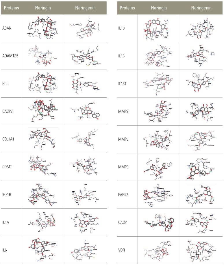
Results: Molecular docking showed that both Nar and NG bind to the selected genes of interest. Semi-quantitative RT-PCR analysis reveals differential gene expression of collagen (COL)9A1, COL9A2, COL9A3, COL11A2, COMT (catechol-O-methyltransferase), and THBS2 (thrombospondin 2); up regulation of ACAN (aggrecan), COL1A1, COL11A1, interleukin (IL)6, IL10, IL18R1, IL18RAP, metalloprotease (MMP)2, MMP3, MMP9, ADAMTS5 (a disintegrin and metalloproteinase with thrombospondin motifs 5), IGF1R (insulin-like growth factor type 1 receptor), SPARC (secreted protein acidic and cysteine rich), PARK2 (parkin), VDR (vitamin D receptor), and BCL2 (B-cell lymphoma 2); down regulation of IL1A, CASP3 (caspase 3), and nine genes with predetermined concentrations of Nar and NG.
Conclusions: The present study evaluated the anti-inflammatory and regenerative efficiencies of Nar and NG in degenerated human NP cells. Altered gene expressions of cytokines, metalloproteases, extracellular proteins, apoptotic genes were dose responsive. The molecular docking (in silico) studies showed effective binding of these native ligands (Nar and NG) with genes identified as potent inhibitors of inflammation. Thus, these natural flavonoids could serve as anti-inflammatory agents in the treatment of low back pain and sciatica.
Keywords: Flavonoids; Inflammation; Intervertebral disc degeneration; Naringin and naringenin; Nucleus pulposus.
Conflict of interest statement
No potential conflict of interest relevant to this article was reported.
Figures


Similar articles
-
Naringin Protects Against Interleukin 1β (IL-1β)-Induced Human Nucleus Pulposus Cells Degeneration via Downregulation Nuclear Factor kappa B (NF-κB) Pathway and p53 Expression.Med Sci Monit. 2019 Dec 25;25:9963-9972. doi: 10.12659/MSM.918597. Med Sci Monit. 2019. PMID: 31927560 Free PMC article.
-
Therapeutic effects of naringin on degenerative human nucleus pulposus cells for discogenic low back pain.Spine J. 2016 Oct;16(10):1231-1237. doi: 10.1016/j.spinee.2016.05.007. Epub 2016 May 18. Spine J. 2016. PMID: 27208552
-
Anti-inflammatory effects of interleukin-4 on intervertebral disc cells.Spine J. 2020 Jan;20(1):60-68. doi: 10.1016/j.spinee.2019.06.025. Epub 2019 Jun 29. Spine J. 2020. PMID: 31265894
-
Genetic Predictors of Early-Onset Spinal Intervertebral Disc Degeneration: Part One of Two.Cureus. 2021 May 22;13(5):e15182. doi: 10.7759/cureus.15182. Cureus. 2021. PMID: 34178503 Free PMC article. Review.
-
Differentiation of Pluripotent Stem Cells into Nucleus Pulposus Progenitor Cells for Intervertebral Disc Regeneration.Curr Stem Cell Res Ther. 2019;14(1):57-64. doi: 10.2174/1574888X13666180918095121. Curr Stem Cell Res Ther. 2019. PMID: 30227822 Review.
Cited by
-
The Anti-Inflammatory and Cytoprotective Efficiency of Curvularin, a Fungal Macrolactone against Lipopolysaccharide-Induced Inflammatory Response in Nucleus Pulposus Cells: An In Vitro Study.Asian Spine J. 2021 Apr;15(2):143-154. doi: 10.31616/asj.2019.0285. Epub 2020 Apr 8. Asian Spine J. 2021. PMID: 32252191 Free PMC article.
-
Exploring the potential anti-apoptotic effects of traditional Chinese medicine in intervertebral disc degeneration: mechanisms and therapeutic prospects.Front Physiol. 2025 Aug 4;16:1617215. doi: 10.3389/fphys.2025.1617215. eCollection 2025. Front Physiol. 2025. PMID: 40832135 Free PMC article. Review.
-
Molecular Mechanisms of Intervertebral Disc Degeneration: Significance of SPARC Gene Expression.J Musculoskelet Neuronal Interact. 2025 Jun 1;25(2):239-247. doi: 10.22540/JMNI-25-239. J Musculoskelet Neuronal Interact. 2025. PMID: 40452200 Free PMC article.
-
Chrysin attenuates intervertebral disk degeneration via dual inhibition of matrix metalloproteinases and senescence: integrated network pharmacology, molecular docking, and experimental validation.Front Med (Lausanne). 2025 May 12;12:1593317. doi: 10.3389/fmed.2025.1593317. eCollection 2025. Front Med (Lausanne). 2025. PMID: 40421294 Free PMC article.
-
An update on animal models of intervertebral disc degeneration and low back pain: Exploring the potential of artificial intelligence to improve research analysis and development of prospective therapeutics.JOR Spine. 2023 Jan 30;6(1):e1230. doi: 10.1002/jsp2.1230. eCollection 2023 Mar. JOR Spine. 2023. PMID: 36994457 Free PMC article. Review.
References
-
- Li Y, Zhu J, Gao C, Peng B. Vitamin D receptor (VDR) genetic polymorphisms associated with intervertebral disc degeneration. J Genet Genomics. 2015;42:135–40. - PubMed
-
- Annunen S, Paassilta P, Lohiniva J, et al. An allele of COL9A2 associated with intervertebral disc disease. Science. 1999;285:409–12. - PubMed
-
- Seki S, Kawaguchi Y, Mori M, et al. Association study of COL9A2 with lumbar disc disease in the Japanese population. J Hum Genet. 2006;51:1063–7. - PubMed
-
- Ito S, Kimura K, Haneda M, Ishida Y, Sawada M, Isobe K. Induction of matrix metalloproteinases (MMP3, MMP12 and MMP13) expression in the microglia by amyloid-beta stimulation via the PI3K/Akt pathway. Exp Gerontol. 2007;42:532–7. - PubMed
-
- Mashayekhi F, Shafiee G, Kazemi M, Dolati P. Lumbar disk degeneration disease and aggrecan gene polymorphism in northern Iran. Biochem Genet. 2010;48:684–9. - PubMed
LinkOut - more resources
Full Text Sources
Research Materials
Miscellaneous
