. 2019 Oct 24;146(20):dev181750.
doi: 10.1242/dev.181750.

Muscle-selective RUNX3 dependence of sensorimotor circuit development

Affiliations

Muscle-selective RUNX3 dependence of sensorimotor circuit development

Yiqiao Wang et al. Development. .

Abstract

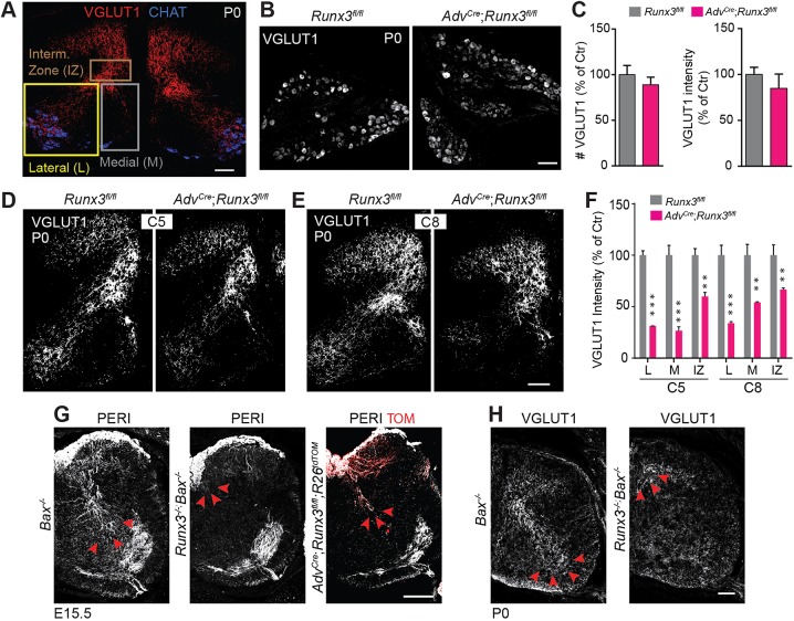
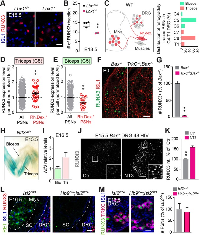
The control of all our motor outputs requires constant monitoring by proprioceptive sensory neurons (PSNs) that convey continuous muscle sensory inputs to the spinal motor network. Yet the molecular programs that control the establishment of this sensorimotor circuit remain largely unknown. The transcription factor RUNX3 is essential for the early steps of PSNs differentiation, making it difficult to study its role during later aspects of PSNs specification. Here, we conditionally inactivate Runx3 in PSNs after peripheral innervation and identify that RUNX3 is necessary for maintenance of cell identity of only a subgroup of PSNs, without discernable cell death. RUNX3 also controls the sensorimotor connection between PSNs and motor neurons at limb level, with muscle-by-muscle variable sensitivities to the loss of Runx3 that correlate with levels of RUNX3 in PSNs. Finally, we find that muscles and neurotrophin 3 signaling are necessary for maintenance of RUNX3 expression in PSNs. Hence, a transcriptional regulator that is crucial for specifying a generic PSN type identity after neurogenesis is later regulated by target muscle-derived signals to contribute to the specialized aspects of the sensorimotor connection selectivity.

Keywords: Dorsal root ganglia; Neuronal specification; Neurotrophins; Sensorimotor circuit; Sensory system.


Conflict of interest statement

Competing interestsThe authors declare no competing or financial interests.

Figures

Fig. 1.
Fig. 1.
Loss of cell identity in subgroups of PSNs following conditional targeting of RUNX3 after peripheral innervation. (A) Scheme representing the successive developmental steps of PSNs, which contribute to sensorimotor circuit. Early specification of PSNs (i) is followed by peripheral axonal growth and muscle targeting (circa. E14) (ii). After peripheral innervation, central afferents of PSNs project to the intermediate and then ventral regions of the spinal cord to contact interneurons and motor neurons (iii). (B) Ablation of Runx3 from sensory neurons using AdvCre mice. At E13.5, RUNX3 expression is detectable in TRKC+ neurons with tdTomato (RFP) starting to be expressed in few neurons, while at E15.5, the recombination is fully efficient, all neurons expressing tdTomato and RUNX3 are strongly reduced in number. Scale bar: 50 µm. (C) Quantification of B, showing the recombination efficiency in TRKC+/RUNX3+ in AdvCre;Runx3fl/fl mice (n=3). ***P≤0.001; Student's t-test. Data are mean±s.e.m. (D) Immunostaining for NF200 and RUNX1 on DRG sections from P0 AdvCre;Runx3fl/fl and Runx3fl/fl animals identifies all myelinated sensory neurons (mechanoreceptive and proprioceptive neurons) and a large majority of nociceptive neurons (Lallemend and Ernfors, 2012; Gascon et al., 2010). Scale bar: 50 µm. (E) Quantification of D reveals absence of cell death in DRG neurons in the conditional Runx3 mutants at P0. P>0.05, Student's t-test. Data are mean±s.e.m. (n=3). (F-K) Immunostaining for PSNs markers (F,H,J) and their quantification (G,I,K) in AdvCre;Runx3fl/fl and Runx3fl/fl P0 animals (n=3 per genotype). Scale bars: 100 µm in F; 50 µm in H,J. n=3 per genotype; *P≤0.05; Student's t-test. (G) Data are mean±s.e.m.
Fig. 2.
Fig. 2.
Central afferentation deficit of PSNs after conditional targeting of RUNX3. (A) Representative regions of the spinal cord analyzed. To reveal central afferent terminations of PSNs, we used VGLUT1 immunostaining on cross-sections of spinal cord. The pattern of VGLUT1 reactivity was analyzed in three reference regions: the intermediate zone (IZ), the ventromedial (M) and the ventrolateral (L) regions. Scale bar: 100 µm. (B) VGLUT1 expression in AdvCre; Runx3fl/fl and Runx3fl/fl DRG sections at P0. Scale bar: 50 µm. (C) Quantification of the number of VGLUT1+ neurons per DRG section (left panel) and VGLUT1 intensity per cell (right panel) (from data in B) reveals absence of change in VGLUT1 expression in DRG from AdvCre;Runx3fl/fl mice (n=3). (D,E) Central innervation of PSNs in AdvCre;Runx3fl/fl and Runx3fl/fl mice at C5 (D) and C8 (E), as revealed by VGLUT1 immunostaining. Scale bar: 100 µm. (F) Quantification of the density of VGLUT1 staining in D and E in regions defined in A on one side of the spinal cord reveals deficits in central ingrowth of PSN afferents in conditional Runx3 mutant mice (n=4 per genotype). A greater difference was observed in the lateral and medial (L and M) regions of the ventral spinal cord, which corresponds to the innervation of the MN pools (CHAT+ in A). **P≤0.01, ***P≤0.001; Student's t-test. Data are mean±s.e.m. (n=3). (G) Immunostaining for peripherin (PERI) on spinal cord sections shows complete absence of central PSN afferents in Runx3−/−;Bax−/− compared with AdvCre;Runx3fl/fl mice (see red arrowheads). Scale bar: 100 µm. (H) Similar to G, at P0 VGLUT1 immunostaining on spinal cord sections confirms the absence of central axon growth of PSNs in Runx3−/−;Bax−/− mice (see red arrowheads), a phenotype that differs from AdvCre;Runx3fl/fl mice (see D,E). Scale bar: 100 µm.
Fig. 3.
Fig. 3.
Muscle-selective differential penetrance of central deficits of PSN connectivity in RUNX3 conditional mutants. (A) Experimental scheme describing muscle injection of the antagonistic muscles biceps and triceps, and retrograde labeling of specific MN pools at spinal segments C5-C6 for the biceps or segments C7-T1 for the triceps. (B) Immunostaining of spinal cord cross-sections representing MNs (CHAT+, in blue) traced by the CTB (red) from initial injection in the biceps. PSN synaptic contacts with the MNs are visualized by the VGLUT1+ synaptic bouton (green). Scale bars: 10 µm. (C) Quantification of 38 MNs (from B) in Runx3fl/fl and 39 MNs in AdvCre;Runx3fl/fl. ***P≤0.001; Student's t-test. Data are mean±s.e.m. (n=3). (D) Immunostaining of spinal cord cross-sections representing MNs (CHAT+, in blue) traced by the CTB (red) from initial injection in the triceps. PSN synaptic contacts with the MNs are visualized by the VGLUT1+ synaptic bouton (green). Scale bars: 10 µm. (E) Quantification of a total of 44 MNs (from D) in Runx3fl/fl and 46 MNs in AdvCre;Runx3fl/fl. ***P≤0.001; Student's t-test. Data are mean±s.e.m. (n=3).
Fig. 4.
Fig. 4.
Muscle-specific MSs deficits in conditional Runx3 mutants at birth. (A) Immunostaining for VGLUT1 (for MSs) and myosin on cross-sections from biceps and triceps. (B) Quantification reveals a muscle-selective MS deficiency in AdvCre;Runx3fl/fl with a significant decrease in the numbers of MSs in triceps (n=3 animals). **P≤0.01, ****P≤0.0001; Student's t-test. Data are mean±s.e.m. Scale bar: 100 µm.
Fig. 5.
Fig. 5.
RUNX3 dependence of PSN connectivity correlates with RUNX3 levels and muscle target NT3 levels. (A) Immunostaining for ISL1 and RUNX3 on DRG sections from Lbx1−/− and Lbx1+/+ mice. Scale bar: 50 µm. (B) Quantification of RUNX3+ neurons (from A) in Lbx1−/− (n=2) and their control littermates (n=4) at brachial levels shows significant reduction in the full mutants compared with control animals. **P≤0.01. Data are mean±s.e.m. (C) Experimental design. (Left) Rhodamine dextran (Rh. dex.) injection in specific muscle will retrogradely trace their innervating PSNs in the DRG from E16.5 wild-type animals. The dextran is injected in biceps in one of the forelimbs and in triceps in the contralateral limb. (Right) Distribution within DRG of the PSNs innervating biceps and triceps, showing biceps- and triceps-innervating PSNs located mostly in DRG C5 and C8, respectively (data from 5 animals for triceps and 11 animals for biceps). (D,E) Quantification of RUNX3 expression per cell in retrogradely traced PSNs (as in C) versus all RUNX3+ PSNs after injection of Rh. dex. in triceps (analysis at C8 level, D) or in biceps (analysis at C5 level, E). **P≤0.01; Student's t-test. Data are mean±s.e.m. (single neurons analyzed from five embryos). (F) RUNX3 expression is largely reduced in TrkC−/−;Bax−/− P0 mice: a mouse model of peripheral outgrowth deficits. Scale bar: 100 µm. (G) Quantification of F reveals an almost complete absence of RUNX3 expression in TrkC−/−;Bax−/− P0 mice compared with their control littermates (n=2). **P≤0.01; Student's t-test. Data are mean±s.e.m. (H) X-Gal reaction on E15.5 in Nft3Lacz/+ forelimb embryos show heterogeneous muscle-specific expression of NT3. There is a large difference in NT3 levels between biceps and triceps. (I) Quantification of NT3 mRNA (Nft3) in the biceps (Bic) and triceps (Tri) shows a twofold increase in triceps compared with biceps in control animals (Ctr). P=0.052; Student's t-test (n=2 samples from four animals). Data are mean±s.e.m. (J) Bax−/− mice DRG explants (E15.5) in culture with or without NT3 for 48 h in vitro (HIV) reveal a decreased expression of RUNX3 in the absence of NT3. Scale bar: 100 µm. (K) Quantification of J shows a significant increase of RUNX3 intensity per cell, while the number of positive neurons remains unchanged. **P≤0.01; Student's t-test. Data are mean±s.e.m. (n=3). (L) Immunostaining for ISL1, RET and RUNX3 on spinal cord (SC) sections from Hb9Cre;Islet2DTA mice shows complete absence of MNs at E16.5. Scale bar: 50 µm. (M) Immunostaining for TRKC, RUNX3 and ISL1 on DRG sections from Hb9Cre;Islet2DTA and Islet2DTA E16.5 mice shows no deficits in RUNX3 and TRKC expression in the absence of MNs, confirmed by the quantification of the number of PSNs in Hb9Cre;Islet2DTA and Islet2DTA (n=2, right panel). Scale bar: 20 µm.

Similar articles

Cited by

References

    1. Arber S., Ladle D. R., Lin J. H., Frank E. and Jessell T. M. (2000). ETS gene Er81 controls the formation of functional connections between group Ia sensory afferents and motor neurons. Cell 101, 485-498. 10.1016/S0092-8674(00)80859-4 - DOI - PubMed
    1. Brohmann H., Jagla K. and Birchmeier C. (2000). The role of Lbx1 in migration of muscle precursor cells. Development 127, 437-445. - PubMed
    1. Chen A. I., de Nooij J. C. and Jessell T. M. (2006). Graded activity of transcription factor Runx3 specifies the laminar termination pattern of sensory axons in the developing spinal cord. Neuron 49, 395-408. 10.1016/j.neuron.2005.12.028 - DOI - PubMed
    1. Chou Y.-H., Zheng X., Beachy P. A. and Luo L. (2010). Patterning axon targeting of olfactory receptor neurons by coupled hedgehog signaling at two distinct steps. Cell 142, 954-966. 10.1016/j.cell.2010.08.015 - DOI - PMC - PubMed
    1. Dasen J. S. (2009). Transcriptional networks in the early development of sensory-motor circuits. Curr. Top. Dev. Biol. 87, 119-148. 10.1016/S0070-2153(09)01204-6 - DOI - PubMed

Publication types

MeSH terms