. 2019 Oct 1;10(5):e01516-19.
doi: 10.1128/mBio.01516-19.

Hydrodynamic Shape Changes Underpin Nuclear Rerouting in Branched Hyphae of an Oomycete Pathogen

Affiliations

Hydrodynamic Shape Changes Underpin Nuclear Rerouting in Branched Hyphae of an Oomycete Pathogen

Edouard Evangelisti et al. mBio. .

Abstract

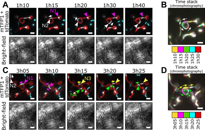
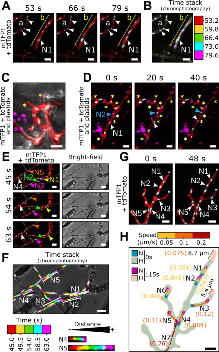
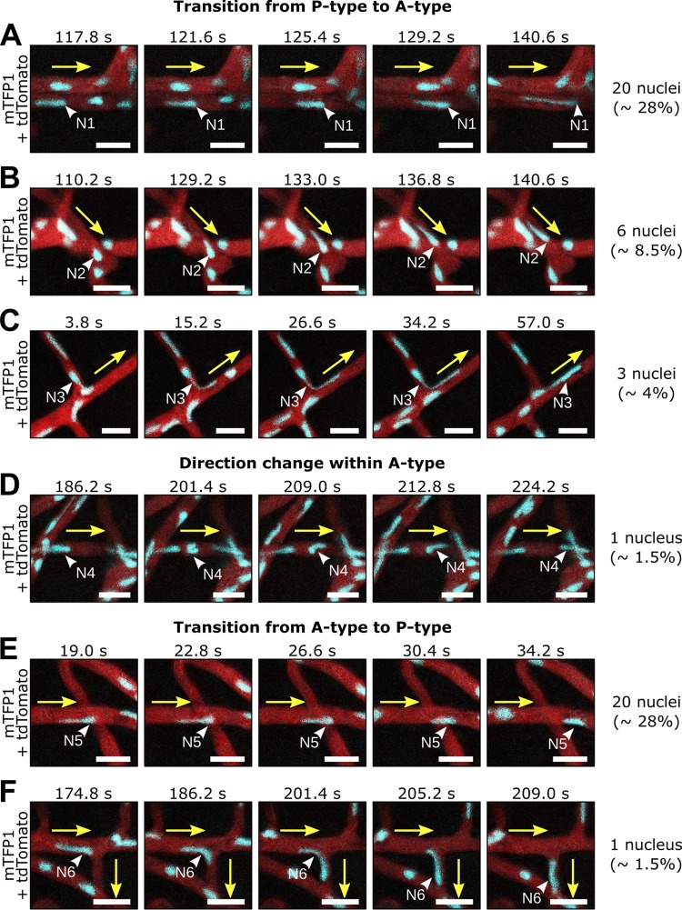
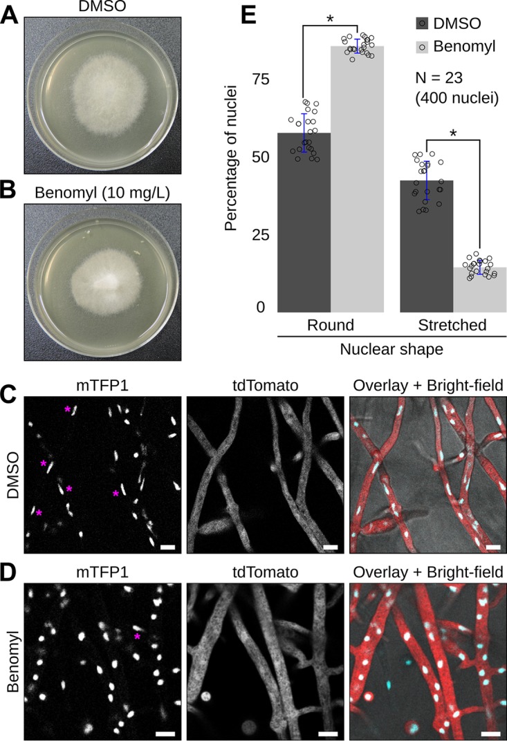
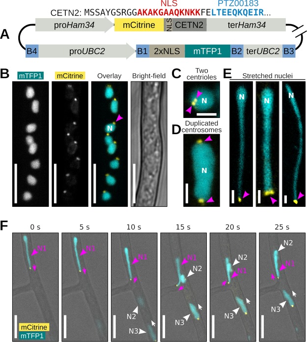
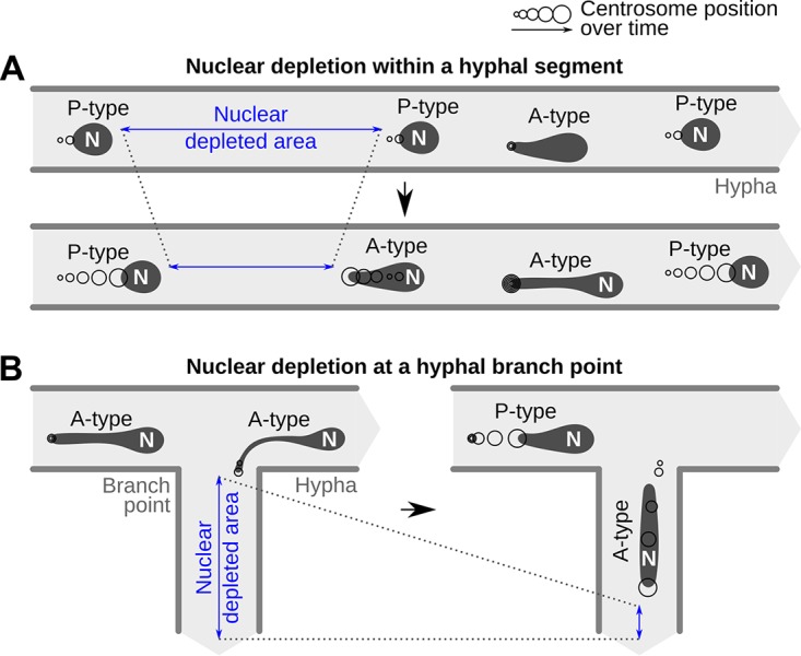
Multinucleate fungi and oomycetes are phylogenetically distant but structurally similar. To address whether they share similar nuclear dynamics, we carried out time-lapse imaging of fluorescently labeled Phytophthora palmivora nuclei. Nuclei underwent coordinated bidirectional movements during plant infection. Within hyphal networks growing in planta or in axenic culture, nuclei either are dragged passively with the cytoplasm or actively become rerouted toward nucleus-depleted hyphal sections and often display a very stretched shape. Benomyl-induced depolymerization of microtubules reduced active movements and the occurrence of stretched nuclei. A centrosome protein localized at the leading end of stretched nuclei, suggesting that, as in fungi, astral microtubule-guided movements contribute to nuclear distribution within oomycete hyphae. The remarkable hydrodynamic shape adaptations of Phytophthora nuclei contrast with those in fungi and likely enable them to migrate over longer distances. Therefore, our work summarizes mechanisms which enable a near-equal nuclear distribution in an oomycete. We provide a basis for computational modeling of hydrodynamic nuclear deformation within branched tubular networks.IMPORTANCE Despite their fungal morphology, oomycetes constitute a distinct group of protists related to brown algae and diatoms. Many oomycetes are pathogens and cause diseases of plants, insects, mammals, and humans. Extensive efforts have been made to understand the molecular basis of oomycete infection, but durable protection against these pathogens is yet to be achieved. We use a plant-pathogenic oomycete to decipher a key physiological aspect of oomycete growth and infection. We show that oomycete nuclei travel actively and over long distances within hyphae and during infection. Such movements require microtubules anchored on the centrosome. Nuclei hydrodynamically adapt their shape to travel in or against the flow. In contrast, fungi lack a centrosome and have much less flexible nuclei. Our findings provide a basis for modeling of flexible nuclear shapes in branched hyphal networks and may help in finding hard-to-evade targets to develop specific antioomycete strategies and achieve durable crop disease protection.

Keywords: Phytophthora palmivora; centrosome; hydrodynamics; nucleus movement; oomycetes.


Figures

FIG 1
FIG 1
Repositioning of daughter nuclei follows division during P. palmivora cyst germination (video 1). (A to D) Time-lapse imaging of germinating cysts from a transgenic P. palmivora strain expressing cytoplasmic tdTomato as well as nucleus-localized mTFP1 (LILI-td-NT) during growth at the surface of N. benthamiana roots. (A) Following first nuclear division, daughter nucleus N1 (magenta arrowhead) migrates toward the tip of the germ tube (white arrow), while the second nucleus, N2 (white arrowhead), remains within the cyst body. (B) Chronophotographic view (time stack) of the previous sequence, showing the successive locations of nucleus N2. Time frames are color coded and overlaid. (C) Nuclear movements following N1 division. Daughter nuclei N3 (yellow arrowhead) and N4 (green arrowhead) move in opposite directions (yellow and green arrows, respectively) and distribute equally within the germ tube. (D) Chronophotographic view (time stack) of the sequence shown in panel C, showing opposite movement of nuclei N3 and N4. Bar, 10 μm.
FIG 2
FIG 2
Appressorium differentiation alters P. palmivora nuclear dynamics in the germ tube. (A and B) Time-lapse imaging of N. benthamiana root infection by a P. palmivora LILI-td-NT strain (video 2). (A) Representative sequence of a successful infection event (video 2A). Arrowheads indicate nuclei. Asterisks indicate migration of the cytoplasm out of the cyst. (B) Representative sequence of an unsuccessful infection event, leading to the differentiation of a second germ tube (indicated by asterisks) opposite the first one (video 2B). Arrowheads indicate nuclei. Bar, 10 μm.
FIG 3
FIG 3
Differential nuclear migration rates occur throughout P. palmivora hyphal network. Time-lapse imaging of P. palmivora LILI-td-NT hyphae infecting an N. benthamiana leaf (A to D, videos 3 and 4) or growing axenically on V8 medium (E to H, videos 5 and 6). (A, video 3) Representative sequence of nuclear movements within infectious P. palmivora hyphae. (B, video 3) Chronophotographic display (time stack) of the sequence shown in panel A. The successive time frames are color coded and overlaid. (C) Representative sequence of P. palmivora haustoria (asterisks) on an N. benthamiana leaf. Arrowheads indicate nuclei. (D, video 4) Representative sequence of nuclear movements within haustoriated P. palmivora hyphae. (E, video 5) Representative sequence of nuclear migration on axenically grown mycelium. (F, video 5) Chronophotographic display of the sequence shown in panel E. (G, video 6) Representative sequence of nuclear movements near hyphal tips. (H) Nuclear trajectories for seven nuclei at the hyphal tip. Average speed is indicated in parentheses. Lines indicate hyphal segments. Arrowheads indicate individual nuclei. N, nuclei; H, hyphae. Bar, 10 μm.
FIG 4
FIG 4
Individual P. palmivora nuclei stretch and move independently of surrounding nuclear flow. (A to F, video 6) Time-lapse imaging of axenically grown P. palmivora LILI-td-NT hyphae containing nuclei transitioning from passive (P-type) to active (A-type) movements (A to D) or, conversely, from A type to P type (E and F). (A) Nucleus slows down and stretches sternward. (B) Nucleus shrinks and starts moving together with the surrounding nuclear flow. (C) Same as panel B, but nucleus enters a branch. (D) Nucleus stretches and moves opposite the surrounding nuclear flow. (E) Same as panel D but entering a branch. (F) Nucleus stretches and moves opposite the surrounding nuclear flow and then tumbles and initiates a similar movement in the opposite direction. Yellow arrows indicate mass nuclear flow. White arrowheads indicate nuclei. Frequencies are given based on a total of 70 nuclei. Bar, 10 μm.
FIG 5
FIG 5
P. palmivora nuclear stretching correlates with rerouting toward nucleus-depleted hyphal segments. (A, video 7) Representative sequence of nuclear movements at a hyphal branch point, highlighting both A-type (nuclei N1 and N2) and P-type (nucleus N3) movements. Arrowheads indicate nuclei. (B to J) Analysis of nuclear speed and trajectories for nuclei N1 to N3. (B, E, and H) Maps of nuclear content in the vicinity of nuclei N1 (B), N2 (E), and N3 (H). Black arrows indicate nuclear movement. (C, F, and I) Nuclear trajectories of N1 (C), N2 (F), and N3 (I). Locations of nuclei N1 to N3 at first (0 s), intermediate (135 s), and last (210 s) time points are shown in blue, red, and magenta, respectively. Nuclear centroids are represented as dots. Arrows indicate instantaneous speed and direction of the movement. Average nucleus speed is indicated on the graph. (D, G, and J) Variation of instantaneous speed with time for nuclei N1 (D), N2 (G), and N3 (J). Average speed in shown as a red dotted line. Representative pictures of nucleus shape over time are shown above the graphs with arrows indicating direction of the movement. Bar, 10 μm.
FIG 6
FIG 6
Antimicrotubule drug Benomyl impairs P. palmivora A-type nuclear movements. (A and B) Growth habit of transgenic P. palmivora LILI-td-NT mycelium growing on V8 agar plates without (dimethyl sulfoxide [DMSO] control) (A) or with (B) 10 mg/liter Benomyl. (C and D) Representative pictures of P. palmivora LILI-td-NT hyphae and nuclei in absence (C, video 8A) or presence (D, video 8B) of 10 mg/liter Benomyl. Bar, 10 μm. (E) Quantification of round (P-type) and stretched (A-type) nuclei within P. palmivora LILI-td-NT hyphae (n = 23, 400 nuclei). Statistical significance was assessed using the Wilcoxon test for paired samples (P < 0.05).
FIG 7
FIG 7
Centrin2 localizes to P. palmivora nuclear stretches. (A) Schematic view of the construct used for dual labeling of nuclei and centrosomes in P. palmivora strain LILI-NT-Ce. Backbone elements are not represented. (B, video 9) Representative picture of mCitrine-labeled Centrin2 (CETN2) within P. palmivora LILI-NT-Ce hyphae. (C) CETN2 localizes in two adjacent dots at the periphery of the nucleus. (D) Centrosome duplication in dividing nuclei. (E) Representative pictures of stretched P. palmivora nuclei (up to 30 μm long), with CETN2 localizing at the tip of the stretched areas. (F) Time-lapse imaging of nuclear movements at hyphal branch point. Nucleus N1 (magenta) follows an A-type trajectory backward to the branch point, while nuclei N2 and N3 follow a P-type trajectory. CETN2 localizes at the tip of the stretched part of nucleus N1, while CETN2 localizes at the back of nuclei N2 and N3. Bar, 2 μm (C to E) or 10 μm (B and F).
FIG 8
FIG 8
Proposed behavior change from passive (P-type) to active (A-type) movements within P. palmivora hyphae. (A) Decrease in nuclear density within a hyphal segment (blue) causes downstream nuclei to anchor until populated by P-type nuclei or to initiate retrograde movement to fill it directly. Anchorage is mediated by the centrosome and results in nuclear stretching. (B) Decrease in nuclear density in a branching hypha (blue) causes nuclei to anchor at the branch point and initiate active movement to enter the branch.

Similar articles

Cited by

References

    1. Morris NR. 2000. Nuclear migration: from fungi to the mammalian brain. J Cell Biol 148:1097. doi:10.1083/jcb.148.6.1097. - DOI - PMC - PubMed
    1. Xiang X. 2018. Nuclear movement in fungi. Semin Cell Dev Biol 82:3–16. doi:10.1016/j.semcdb.2017.10.024. - DOI - PMC - PubMed
    1. Veneault-Fourrey C, Barooah M, Egan M, Wakley G, Talbot NJ. 2006. Autophagic fungal cell death is necessary for infection by the rice blast fungus. Science 312:580–583. doi:10.1126/science.1124550. - DOI - PubMed
    1. Fernandez J, Marroquin-Guzman M, Wilson RA. 2014. Evidence for a transketolase-mediated metabolic checkpoint governing biotrophic growth in rice cells by the blast fungus Magnaporthe oryzae. PLoS Pathog 10:e1004354. doi:10.1371/journal.ppat.1004354. - DOI - PMC - PubMed
    1. Jeon J, Rho H, Kim S, Kim KS, Lee YH. 2014. Role of MoAND1-mediated nuclear positioning in morphogenesis and pathogenicity in the rice blast fungus, Magnaporthe oryzae. Fungal Genet Biol 69:43–51. doi:10.1016/j.fgb.2014.05.002. - DOI - PubMed

Publication types

LinkOut - more resources