. 2019 Oct 1;9(1):14103.
doi: 10.1038/s41598-019-50679-y.

Vitamin D3 regulates apoptosis and proliferation in the testis of D-galactose-induced aged rat model

Affiliations

Vitamin D3 regulates apoptosis and proliferation in the testis of D-galactose-induced aged rat model

Malsawmhriatzuala Jeremy et al. Sci Rep. .

Abstract

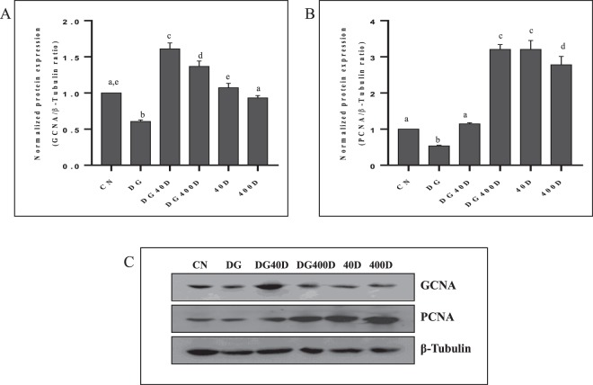
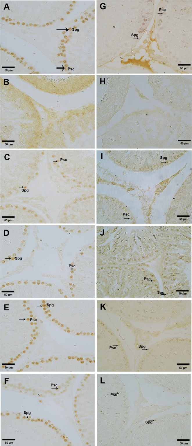
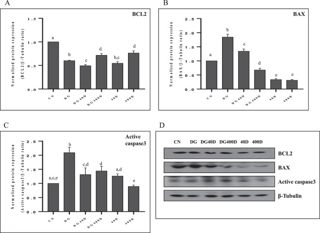
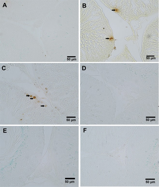
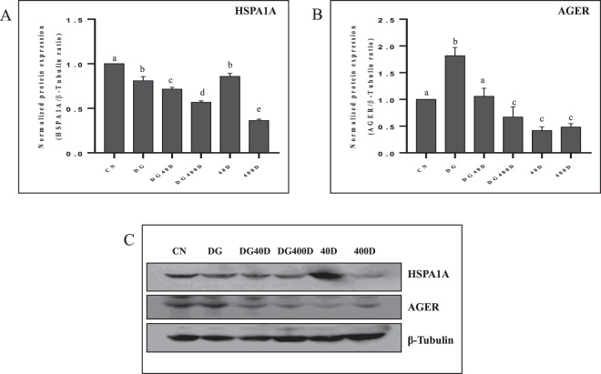
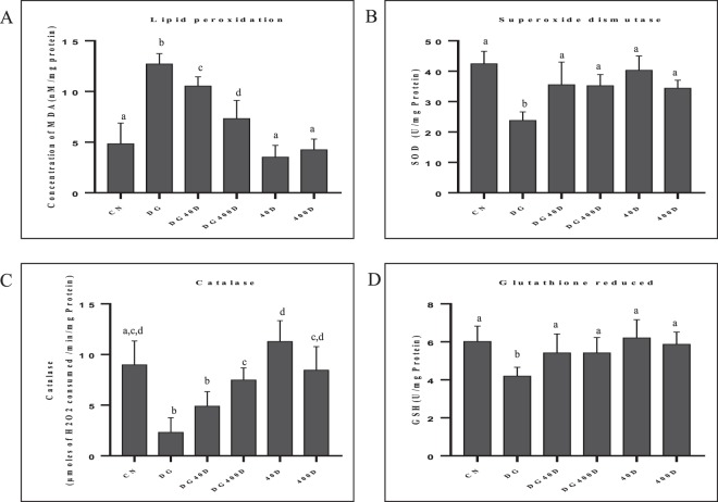
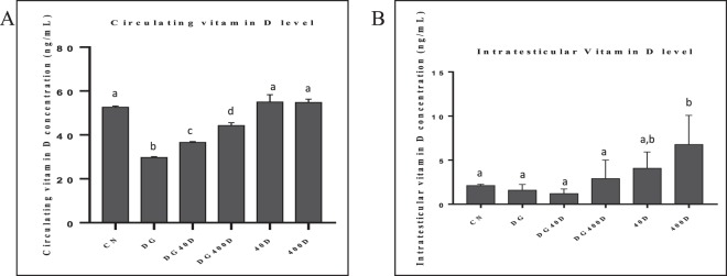
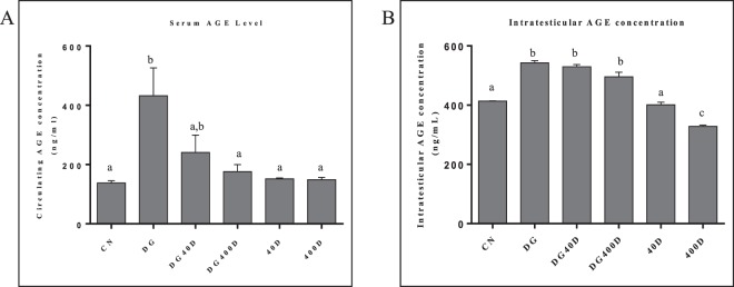
The age-associated imbalances between proliferation and apoptosis lead to impaired spermatogenesis and infertility. The age-associated decline in vitamin D3 levels has been reported and suggested the anti-aging potential of vitamin D3. However, the age-associated decline levels of vitamin D3 has not been studied in relation to the testicular activity. Thus, we investigated the effect of vitamin D3 on the expression of testicular proliferation markers, apoptotic markers, antioxidants system and oxidative stress in a D-gal-induced aged rat model. The present study investigated the levels of vitamin D3 and AGE in serum and testes along with the expression of the AGE-receptor (AGER) in the testis. Vitamin D3 treatment significantly increases cell proliferation and decreases apoptosis in a D-gal-induced aged rat testis. Furthermore, vitamin D3 significantly decreases oxidative stress in aged rat testis by improving the antioxidant defense systems. The expression of AGER was down-regulated by vitamin D3 treatment in aged testis. The circulating and intra-testicular AGE was higher in aged groups, however, only circulating vitamin D3 levels decreased in aged groups. The immunolocalization of VDR showed increased immunostaining in the testis by vitamin D3 treatment. Thus, it can be concluded that vitamin D3 delays testicular senescence by regulating proliferation and apoptosis.


Conflict of interest statement

The authors declare no competing interests.

Figures

Figure 1
Figure 1
Vitamin D3 increased germ cell proliferation in D-gal-induced aged rat testis. (A) Densitometry of Western blots of GCNA protein expression in rat testis. (B) Densitometry of Western blots of PCNA protein expression in rat testis. (C) Representative Western blots of GCNA and PCNA protein expression in rat testis. Values are mean ± SEM (n = 6) and different superscripts show significant (P < 0.05) difference in protein expression. CN Control, DG D-galactose, DG40D D-galactose plus 40 IU/Kg vitamin D3, DG400D D-galactose plus 400 IU/Kg vitamin D3, 40D 40 IU/Kg vitamin D3, 400D 400 IU/Kg vitamin D3 groups. Cropped blots are shown. Uncropped blots are presented in Supplementary Fig. S1.
Figure 2
Figure 2
Effect of vitamin D3 on the localization of GCNA and PCNA in testis. A-F Localization of PCNA also showed its expression in spermatogonia (Spg) and spermatocytes (Psc) arrow) in the testis of control (A), D-gal-induced aged rat testis treated with vitamin D3 at dose of 40 IU/Kg (C), 400 IU/Kg (D), normal rat treated with alone vitamin D3 at dose of 40 IU/Kg (E) and 400 IU/Kg. (F) D-gal-induced aged rat testis showed no immunostaining of PCNA. (B) G-L Localization of GCNA confined to spermatogonia (Spg) and spermatocytes (Psc) (arrow) in the testis of control (G), D-gal-induced aged rat testis treated with vitamin D3 at dose of 40 IU/Kg (I), 400 IU/Kg (J), normal rat treated with alone vitamin D3 at dose of 40 IU/Kg (K) and 400 IU/Kg. (L) D-gal-induced aged rat testis showed no immunostaining of GCNA. (H) The figure shows a magnification of 40x.
Figure 3
Figure 3
Vitamin D3 dependent expression of anti-apoptotic (BCL2) and apoptotic (BAX and active caspase-3) in the testis. (A) Densitometry of Western blots of BCL2 protein expression in rat testis. (B) Densitometry of Western blots of BAX protein expression in rat testis. (C) Densitometry of Western blots of active caspase-3 protein expression in rat testis. (D) Representative Western blots of BCL2, BAX and active caspase-3 protein expression in rat testis. Values are mean ± SEM (n = 6) and different superscripts show significant (P < 0.05) difference in protein expression. CN Control, DG D-galactose, DG40D D-galactose plus 40 IU/Kg vitamin D3, DG400D D-galactose plus 400 IU/Kg vitamin D3, 40D 40 IU/Kg vitamin D3, 400D 400 IU/Kg vitamin D3 groups. Cropped blots are shown. Uncropped blots are presented in Supplementary Fig. S1.
Figure 4
Figure 4
TUNEL immunohistochemical processing of the rat testis reveals the incidence of apoptosis cells in different treatment groups. TUNEL positive germ cells show apoptotic in D-gal-induced aged rat testis (B, arrow). The treatment of vitamin D3 at a dose of 40 IU/Kg to D-gal-induced aged rat also shows a number of TUNEL positive cells, shows apoptosis (C, arrow). However other groups showed no TUNEL positive germ cells in rat testis (A,DF). The figure shows 40X magnifications.
Figure 5
Figure 5
Vitamin D3 modulates the expression of HSP1A1 and AGER in aged rat testis. (A) Densitometry of Western blots of HSP1A1 protein expression in rat testis. (B) Densitometry of Western blots of AGER protein expression in rat testis. (C) Representative Western blots of HSP1A1 and AGER protein expression in rat testis. Values are mean ± SEM (n = 6) and different superscripts show significant (P < 0.05) difference in protein expression. CN Control, DG D-galactose, DG40D D-galactose plus 40 IU/Kg vitamin D3, DG400D D-galactose plus 400 IU/Kg vitamin D3, 40D 40 IU/Kg vitamin D3, 400D 400 IU/Kg vitamin D3 groups. Cropped blots are shown. Uncropped blots are presented in Supplementary Fig. S1.
Figure 6
Figure 6
Vitamin D3 mediated regulation of oxidative stress and antioxidant systems (SOD, catalase and GSH) in rat testis. (A) Oxidative stress measured as MDA levels and values are expressed nM/mg of protein. (B) SOD activity expressed in U/mg of protein. (C) Catalase activity expressed as µmole of H2O2 consumed/min/mg of protein. (D) Reduced glutathione (GSH) expressed as U/mg of protein. Values are mean ± SEM (n = 6) and different superscripts show significant (P < 0.05) difference. CN Control, DG D-galactose, DG40D D-galactose plus 40 IU/Kg vitamin D3, DG400D D-galactose plus 400 IU/Kg vitamin D3, 40D 40 IU/Kg vitamin D3, 400D 400 IU/Kg vitamin D3 groups.
Figure 7
Figure 7
Effect of vitamin D3 on circulating and intra-testicular vitamin D3 levels. (A) Circulating vitamin D3 levels showed significant decreased (P < 0.05) in D-gal-induced aged rat. (B) Intra-testicular vitamin D3 levels showed significant (P < 0.05) increased in the testis of normal rat treated with vitamin D3 compared to other groups. Values are mean ± SEM (n = 6) and different superscripts show significant (P < 0.05) difference. CN Control, DG D-galactose, DG40D D-galactose plus 40 IU/Kg vitamin D3, DG400D D-galactose plus 400 IU/Kg vitamin D3, 40D 40 IU/Kg vitamin D3, 400D 400 IU/Kg vitamin D3 groups.
Figure 8
Figure 8
Effect of vitamin D3 on circulating and intra-testicular AGE levels. (A) Circulating AGE levels showed significant increased (P < 0.05) in D-gal-induced aged rat group and D-gal aged art treated with vitamin D3 40 IU/Kg groups compared to other groups (DG and DG40D). (B) Intra-testicular AGE levels showed significant (P < 0.05) increased in the testis of DG, DG40D and DG400D compared to other groups. Values are mean ± SEM (n = 6) and different superscripts show significant (P < 0.05) difference. CN Control, DG D-galactose, DG40D D-galactose plus 40 IU/Kg vitamin D3, DG400D D-galactose plus 400 IU/Kg vitamin D3, 40D 40 IU/Kg vitamin D3, 400D 400 IU/Kg vitamin D3 groups.
Figure 9
Figure 9
A-F Effect of vitamin D3 on the localization of VDR in the testis of different groups. Control rat showed localization of VDR in Leydig cells (L), Sertoli cells, Spermatogonia (Spg) and Primary spermatocytes (Psc). (A) The testis of D-gal-induced aged rat showed no immunostaining of VDR. (B) The treatment of vitamin D3 resume the localization of VDR in the testis. (C,D) The alone treatment of vitamin D3 to normal rat also showed distinct localization of VDR in the testis. (E,F) The figure showed 40X magnifications.

References

    1. Blomberg Jensen M, et al. Vitamin D receptor and vitamin D metabolizing enzymes are expressed in the human male reproductive tract. Hum. Reprod. 2010;25:1303–1311. doi: 10.1093/humrep/deq024. - DOI - PubMed
    1. Shahrokhi SZ, Ghaffari F, Kazerouni F. Role of vitamin D in female reproduction. Clin. Chim. Acta. 2016;455:33–38. doi: 10.1016/j.cca.2015.12.040. - DOI - PubMed
    1. Lanske B, Razzaque MS. Vitamin D and aging: old concepts and new insights. J. Nutr. Biochem. 2007;18:771–777. doi: 10.1016/j.jnutbio.2007.02.002. - DOI - PMC - PubMed
    1. Boisen IM, et al. Possible influence of vitamin D on male reproduction. J. Steroid Biochem. Mol. Biol. 2017;173:215–222. doi: 10.1016/j.jsbmb.2016.09.023. - DOI - PubMed
    1. Kwiecinski GG, Petrie GI, DeLuca HF. Vitamin D is necessary for reproductive functions of the male rat. J. Nutr. 1989;119:741–744. doi: 10.1093/jn/119.5.741. - DOI - PubMed

Publication types