. 2019 Oct 1;9(1):14095.
doi: 10.1038/s41598-019-50410-x.

Diversity of vaginal microbiome and metabolome during genital infections

Affiliations

Diversity of vaginal microbiome and metabolome during genital infections

Camilla Ceccarani et al. Sci Rep. .

Abstract

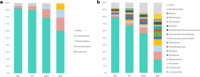
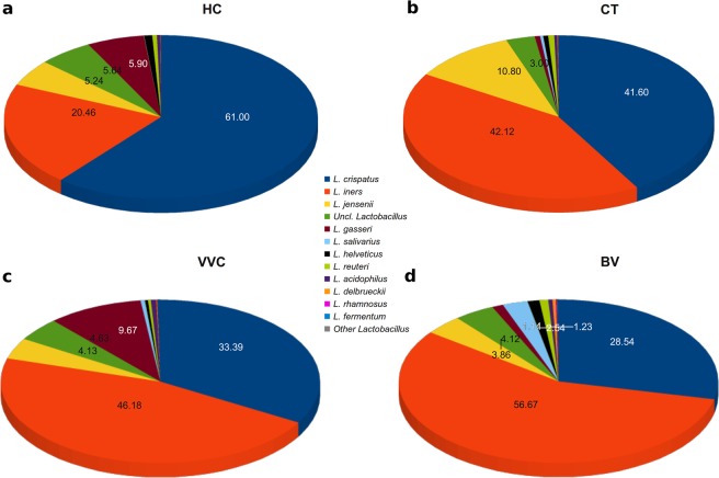
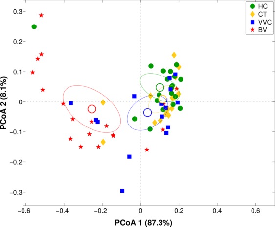
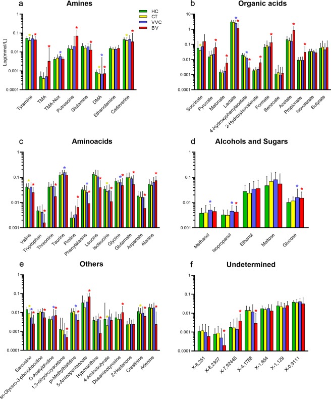
We characterized the vaginal ecosystem during common infections of the female genital tract, as vulvovaginal candidiasis (VVC, n = 18) and Chlamydia trachomatis infection (CT, n = 20), recruiting healthy (HC, n = 21) and bacterial vaginosis-affected (BV, n = 20) women as references of eubiosis and dysbiosis. The profiles of the vaginal microbiome and metabolome were studied in 79 reproductive-aged women, by means of next generation sequencing and proton based-nuclear magnetic resonance spectroscopy. Lactobacillus genus was profoundly depleted in all the genital infections herein considered, and species-level analysis revealed that healthy vaginal microbiome was dominated by L. crispatus. In the shift from HC to CT, VVC, and BV, L. crispatus was progressively replaced by L. iners. CT infection and VVC, as well as BV condition, were mainly characterised by anaerobe genera, e.g. Gardnerella, Prevotella, Megasphaera, Roseburia and Atopobium. The changes in the bacterial communities occurring during the genital infections resulted in significant alterations in the vaginal metabolites composition, being the decrease of lactate a common marker of all the pathological conditions. In conclusion, according to the taxonomic and metabolomics analysis, we found that each of the four conditions is characterized by a peculiar vaginal microbiome/metabolome fingerprint.


Conflict of interest statement

The authors declare no competing interests.

Figures

Figure 1
Figure 1
Structure of the vaginal microbiota. Microbial composition of vaginal swabs of healthy (HC), C. trachomatis (CT), vulvo-vaginal candidiasis (VVC) and bacterial vaginosis (BV) positive women was analysed. (a) Boxplots of Shannon index (α-diversity). (b) Principal Coordinates Analysis (PCoA) plot based on unweighted Unifrac distance (β-diversity). Each point corresponds to a sample. For each experimental class, the SEM-based confidence ellipse around the centroid is depicted. The first two components of the variance are represented.
Figure 2
Figure 2
Taxonomic composition of the vaginal microbiota. Stacked bar charts of taxonomy relative abundances at (a) phylum and (b) genus level for healthy (HC), C. trachomatis (CT), vulvo-vaginal candidiasis (VVC) and bacterial vaginosis (BV) positive subjects. Only phyla and genera present at relative abundances >1% in at least 20% (i.e.: ≥16 samples) are reported. Remaining taxa are grouped in the “Other” category.
Figure 3
Figure 3
Species-level characterization of Lactobacillus genus. Sequences classified within Lactobacillus genus were characterized down to species level by classifying them against a custom reference database. Proportions are relative to the total amount of Lactobacillus genus in each group. Only the twelve most-abundant species are represented. Less abundant Lactobacillus species are grouped under “Other Lactobacillus”. Per-sample relative abundances were averaged in each experimental category: (a) healthy (HC), (b) C. trachomatis (CT), (c) vulvo-vaginal candidiasis (VVC) and (d) bacterial vaginosis (BV) positive women.
Figure 4
Figure 4
Vaginal metabolome analysis. Principal Coordinates Analysis (PCoA) on Bray-Curtis distance showing the separation of the samples according to metabolite profile as obtained from 1H-NMR analysis of vaginal samples from healthy (HC), C. trachomatis (CT), vulvo-vaginal candidiasis (VVC) and bacterial vaginosis (BV) positive women. The first and second components of the total variance are shown.
Figure 5
Figure 5
Metabolite concentration in vaginal samples. Bar plots showing metabolites concentrations per vaginal samples from healthy (HC), C. trachomatis (CT), vulvo-vaginal candidiasis (VVC) and bacterial vaginosis (BV) positive women. Each bar represents the median metabolite concentration (mmol/L) across replicates per experimental group. Error bars show inter-quartile range (i.e.: 75th–25th quantile). Metabolites are grouped according to biochemical type (panels a–f). Stars indicate significant differences vs HC group.
Figure 6
Figure 6
Correlation between metabolome and microbiota. Heatmap showing the Spearman’s correlation coefficient between metabolites concentration and the relative abundances of the main bacterial genera. Only groups present at >1% of relative abundance in at least one sample were considered. Metabolite and microbial data were clustered using Pearson’s correlation metric and average linkage.

References

    1. Ravel J, et al. Vaginal microbiome of reproductive-age women. Proc. Natl. Acad. Sci. 2011;108:4680–4687. doi: 10.1073/pnas.1002611107. - DOI - PMC - PubMed
    1. Younes JA, et al. Women and their microbes: the unexpected friendship. Trends Microbiol. 2018;26:16–32. doi: 10.1016/j.tim.2017.07.008. - DOI - PubMed
    1. Parolin C, et al. Isolation of vaginal lactobacilli and characterization of anti-candida activity. PLoS One. 2015;10:1–17. doi: 10.1371/journal.pone.0131220. - DOI - PMC - PubMed
    1. Calonghi N, et al. Interaction of vaginal Lactobacillus strains with HeLa cells plasma membrane. Benef. Microbes. 2017;8:625–633. doi: 10.3920/BM2016.0212. - DOI - PubMed
    1. Parolin C, et al. Lactobacillus crispatus BC5 interferes with Chlamydia trachomatis infectivity through integrin modulation in cervical cells. Front. Microbiol. 2018;9:e2630. doi: 10.3389/fmicb.2018.02630. - DOI - PMC - PubMed