. 2019 Oct 15;8(20):e012338.
doi: 10.1161/JAHA.119.012338. Epub 2019 Oct 2.

Targeting Extracellular Heat Shock Protein 70 Ameliorates Doxorubicin-Induced Heart Failure Through Resolution of Toll-Like Receptor 2-Mediated Myocardial Inflammation

Affiliations

Targeting Extracellular Heat Shock Protein 70 Ameliorates Doxorubicin-Induced Heart Failure Through Resolution of Toll-Like Receptor 2-Mediated Myocardial Inflammation

Peng Liu et al. J Am Heart Assoc. .

Abstract

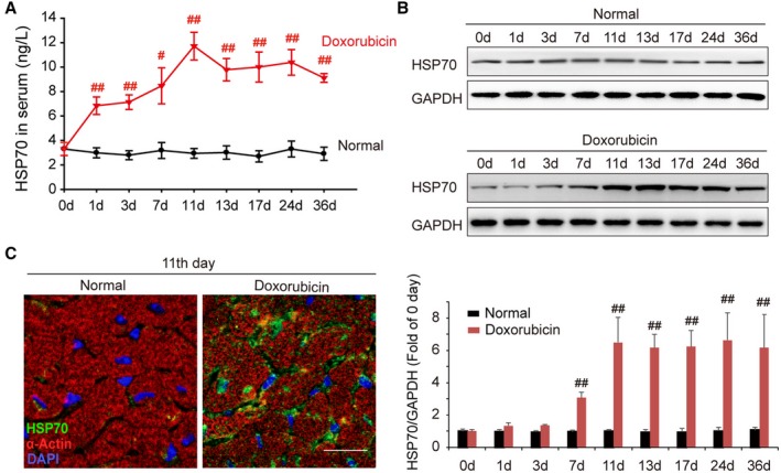
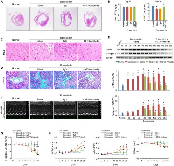
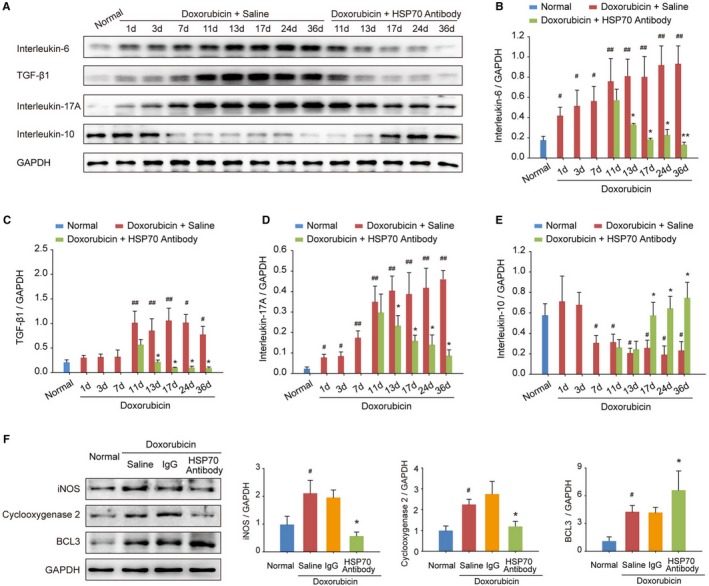
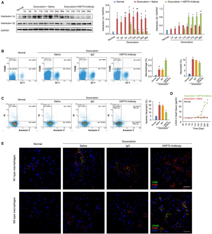
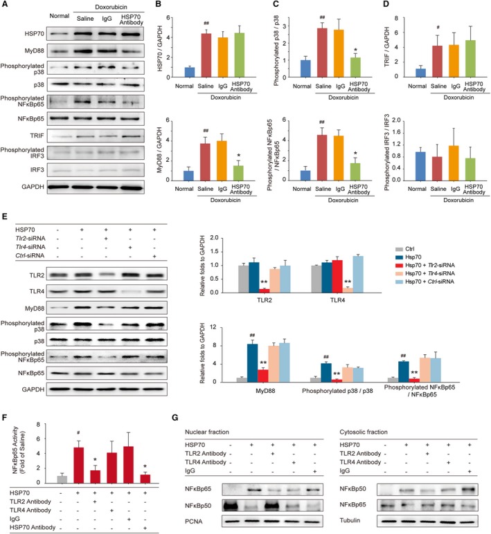
Background Heart failure (HF) is one of the most significant causes of morbidity and mortality for the cardiovascular risk population. We found previously that extracellular HSP70 (heat shock protein) is an important trigger in cardiac hypertrophy and fibrosis, which are associated with the development of heart dysfunction. However, the potential role of HSP70 in response to HF and whether it could be a target for the therapy of HF remain unknown. Methods and Results An HF mouse model was generated by a single IP injection of doxorubicin at a dose of 15 mg/kg. Ten days later, these mice were treated with an HSP70 neutralizing antibody for 5 times. We observed that doxorubicin treatment increased circulating HSP70 and expression of HSP70 in myocardium and promoted its extracellular release in the heart. Blocking extracellular HSP70 activity by its antibody significantly ameliorated doxorubicin-induced left ventricular dilation and dysfunction, which was accompanied by a significant inhibition of cardiac fibrosis. The cardioprotective effect of the anti-HSP70 antibody was largely attributed to its ability to promote the resolution of myocardial inflammation, as evidenced by its suppression of the toll-like receptor 2-associated signaling cascade and modulation of the intracellular distribution of the p50 and p65 subunits of nuclear factor-κB. Conclusions Extracellular HSP70 serves as a noninfectious inflammatory factor in the development of HF, and blocking extracellular HSP70 activity may provide potential therapeutic benefits for the treatment of HF.

Keywords: cardiac dysfunction; cardiac remodeling; damage‐associated molecular patterns; nuclear factor‐κB; toll‐like receptor.


Figures

Figure 1
Figure 1
Doxorubicin elevates the level of HSP (heat shock protein) 70 in serum and myocardium. A, Time course of the content of HSP70 in mouse serum after intraperitoneal administration of doxorubicin. Data are the mean±SEM of 3 assays (n=6–8/group). # P<0.05, ## P<0.01, compared with normal mice. B, At the indicated intervals, the expression of HSP70 in the heart was analyzed by Western blot assay. Representative immunoblots and the ratio of HSP70/GAPDH are presented. Data are the mean±SEM of 3 assays. ## P<0.01, compared with normal mice before doxorubicin treatment. C, Localization of HSP70 in cardiac tissue was detected by confocal microscopy on day 11 after the treatment with doxorubicin (green, HSP70; red, α‐actin; blue, nuclei; bar=20 μm). DAPI indicates 4′,6‐diamidino‐2‐phenylindole.
Figure 2
Figure 2
Blocking extracellular HSP (heat shock protein) 70 activity attenuates doxorubicin‐induced cardiac remodeling and dysfunction. A, Blocking extracellular HSP70 with a neutralizing antibody attenuated doxorubicin‐induced ventricular dilatation (global heart section). B, The ratios of heart weight/body weight (HW/BW) and HW/tibia length (HW/TL) are presented (n=6–8/group). # P<0.05, compared with normal mice; *P<0.05, compared with doxorubicin‐treated mice. C, Blocking extracellular HSP70 inhibited the recruitment of inflammatory cells (bar=50 μm), as indicated by hematoxylin‐eosin (H&E) staining of the heart sections. D, Representative photomicrographs of Masson's trichrome staining of the heart sections for cardiac fibrosis evaluation (bar=50 μm). E, Expression of α‐smooth muscle actin (α‐SMA) and collagen I was assayed by Western blot analysis. The expression ratio of the indicated protein to GAPDH from 3 independent experiments is presented. # P<0.05, compared with normal mice; *P<0.05, compared with doxorubicin‐treated mice. F, Representative images of left ventricular (LV) M‐mode echocardiograms (n=6–8/group). Cardiac functional (G) and structural (H) parameters were measured by echocardiography analysis. Data are mean±SEM (n=6–8/group). # P<0.05, compared with normal mice; *P<0.05, compared with doxorubicin‐treated mice. LVAWd indicates LV diastolic anterior wall thickness; LVIDd, end‐diastolic LV internal diameter; LVIDs, end‐systolic LV internal diameter.
Figure 3
Figure 3
Blocking extracellular HSP (heat shock protein) 70 restricts doxorubicin‐induced myocardial inflammation. A through E, At the indicated time points, the protein levels of interleukin‐6, transforming growth factor (TGF)‐β1, interleukin‐17A, and interleukin‐10 in heart tissue homogenates were detected by Western blot analysis. In a time‐dependent manner, HSP70 neutralizing antibody treatment inhibited the increase in the levels of interleukin‐6 (B), TGF‐β1 (C), and interleukin‐17A (D) but upregulated the level of interleukin‐10 (E) in the myocardium. Representative immunoblots and the ratio of the indicated protein to GAPDH are presented. Data are the mean±SEM of 3 assays. # P<0.05, ## P<0.01, compared with normal mice; *P<0.05, **P<0.01, compared with doxorubicin‐treated mice. F, The expression of the proinflammatory proteins inducible NO synthase (iNOS) and cyclooxygenase 2 as well as the anti‐inflammatory protein B‐cell lymphoma 3 (BCL3) in heart tissue was examined by Western blot analysis at the experimental end point. Representative immunoblots and the ratio of the indicated protein to GAPDH are presented. Data are the mean±SEM of 3 assays. # P<0.05, compared with normal mice; *P<0.05, compared with doxorubicin‐treated mice.
Figure 4
Figure 4
Antagonism of extracellular HSP (heat shock protein) 70 promotes resolution of inflammation in the heart tissue of doxorubicin‐treated mice. A, The time‐dependent expressions of interleukin‐1α and interleukin‐1β in the mouse myocardium after administration of doxorubicin and/or HSP70 antibody. Representative immunoblots and the ratio of the indicated protein to GAPDH are presented. B, The heart single‐cell suspensions were prepared, and the Gr‐1–positive neutrophils and F4/80‐positive macrophages were determined by flow cytometry analysis. Representative scattergrams and the percentage of neutrophils and macrophages are presented. C, The apoptosis of neutrophils was determined by the annexin V and propidium iodide (PI) staining, followed by flow cytometry. Representative scattergrams and percentage of apoptotic neutrophils are presented. D, At the indicated time points, the contents of lipoxin A4 (LXA4) in heart tissue homogenates were detected by ELISA analysis. All data are the mean±SEM of 3 assays. # P<0.05, ## P<0.01, compared with normal mice; *P<0.05, **P<0.01, compared with doxorubicin‐treated mice. E, Recruitment and infiltration of M1‐type macrophages (F4/80+CD86+) and M2‐type macrophages (F4/80+CD206+) in myocardium were detected by confocal microscopy analysis on day 36 after the treatment with doxorubicin (bar=50 μm).
Figure 5
Figure 5
Extracellular HSP (heat shock protein) 70 activates nuclear factor (NF)‐κBp65 and modulates the distribution of the NF‐κB p50 and p65 subunits in a toll‐like receptor 2 (TLR2)–dependent manner. A, Expressions of HSP70, MyD88, phosphorylated p38, p38, phosphorylated NF‐κBp65, NF‐κBp65, toll like receptor adaptor molecule 1 (TRIF), phosphorylated interferon regulatory factor 3 (IRF3), and IRF3 in the mouse myocardium were detected by Western blot analysis. Neutralizing extracellular HSP70 inhibited the MyD88–p38–NF‐κB pathway (B and C) but did not affect the TRIF‐IRF3 pathway (D). Representative immunoblots and the ratio of the indicated protein to GAPDH are presented. # P<0.05, ## P<0.01, compared with normal mice; *P<0.05, **P<0.01, compared with doxorubicin‐treated mice. E, Tlr2–small interfering RNA (siRNA)–, Tlr4‐siRNA–, or Ctrl‐siRNA–transfected H9C2 cells were treated with 100 ng/mL HSP70 recombinant protein for 24 hours; and the TLR2, TLR4, MyD88, phosphorylated p38, p38, phosphorylated NF‐κBp65, and NF‐κBp65 were detected by Western blot analysis. Representative immunoblots and the ratio of the indicated protein to GAPDH are presented. ## P<0.01, compared with untreated cells; **P<0.01, compared with HSP70‐treated cells. F and G, TLR2 antibody, TLR4 antibody, or IgG preincubated H9C2 cells were treated with HSP70 recombinant protein. F, The phosphorylation of NF‐κBp65 in cell homogenates was detected by ELISA analysis. # P<0.05, compared with untreated cells; *P<0.05, compared with HSP70‐treated cells. G, Nuclear and cytosolic extracts were subjected to Western blot analysis with antibodies to the NF‐κB subunits p50 and p65. PCNA and tubulin were used as internal controls for the nuclear and cytosolic fractions, respectively. Representative immunoblots of 3 independent assays are presented.

Similar articles

Cited by

References

    1. Braunwald E. The war against heart failure: the Lancet lecture. Lancet. 2015;385:812–824. - PubMed
    1. Mebazaa A, Yilmaz MB, Levy P, Ponikowski P, Peacock WF, Laribi S, Ristic AD, Lambrinou E, Masip J, Riley JP, McDonagh T, Mueller C, deFilippi C, Harjola VP, Thiele H, Piepoli MF, Metra M, Maggioni A, McMurray JJ, Dickstein K, Damman K, Seferovic PM, Ruschitzka F, Leite‐Moreira AF, Bellou A, Anker SD, Filippatos G. Recommendations on pre‐hospital and early hospital management of acute heart failure: a consensus paper from the Heart Failure Association of the European Society of Cardiology, the European Society of Emergency Medicine and the Society of Academic Emergency Medicine–short version. Eur Heart J. 2015;36:1958–1966. - PubMed
    1. Zhang Y, Bauersachs J, Langer HF. Immune mechanisms in heart failure. Eur J Heart Fail. 2017;19:1379–1389. - PubMed
    1. Van Linthout S, Tschope C. Inflammation—cause or consequence of heart failure or both? Curr Heart Fail Rep. 2017;14:251–265. - PMC - PubMed
    1. Mann DL. Innate immunity and the failing heart: the cytokine hypothesis revisited. Circ Res. 2015;116:1254–1268. - PMC - PubMed

Publication types

MeSH terms