Case Reports
. 2019 Aug;40(4):369-375.
doi: 10.1080/13816810.2019.1660382. Epub 2019 Sep 6.

Modification of the PROM1 disease phenotype by a mutation in ABCA4

Affiliations
Case Reports

Modification of the PROM1 disease phenotype by a mutation in ABCA4

Winston Lee et al. Ophthalmic Genet. 2019 Aug.

Abstract

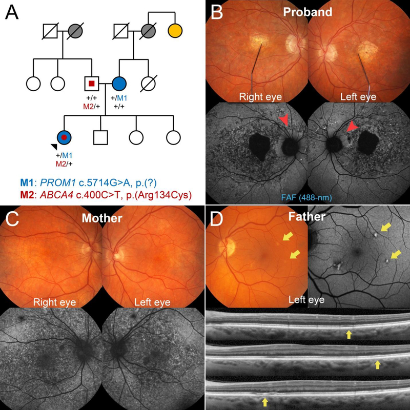
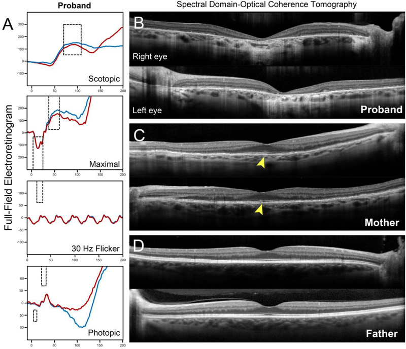
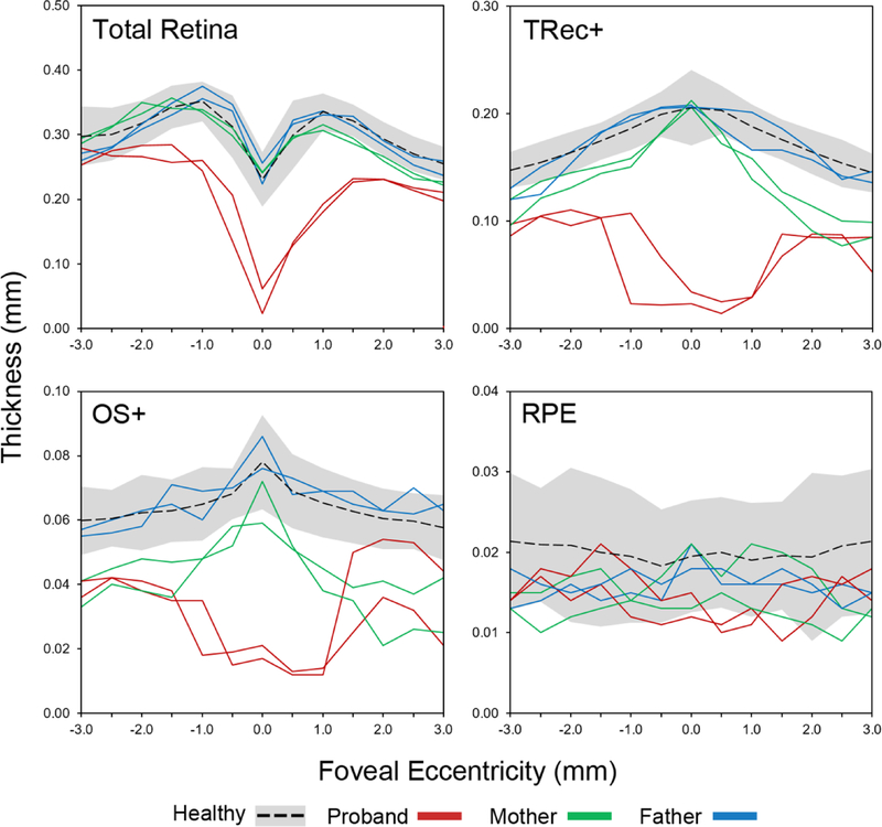
Background: The extensive phenotypic heterogeneity of monogenic diseases can be largely traced to intragenic variation; however, recent advances in clinical detection and gene sequencing have uncovered the emerging role of non-allelic variation (i.e. genetic trans-modifiers) in shaping disease phenotypes. Identifying these associations are not only of significant diagnostic value, but also provides scientific insight into the expanded molecular etiology of rare diseases. This reports describes the discordant clinical manifestation of a family segregating mutations in ABCA4 and PROM1. Methods: Three patients across a two generation family underwent multimodal imaging and functional testing of the retina including color photography, fundus autofluorescence (AF), spectral domain-optical coherence tomography (SD-OCT) and full-field electroretinography (ffERG). Genetic characterization was carried out by direct Sanger and whole exome sequencing. Results: Clinical examination revealed similar retinal degenerative phenotypes in the proband and her mother. Despite being younger, the proband's phenotype was more advanced and exhibited additional features related to Stargardt disease not found in the mother. Whole exome sequencing identified a pathogenic missense variant in PROM1, c.400C > T, p.(Arg134Cys), as the underlying cause of retinal disease in both the proband and mother. Sequencing of the ABCA4 locus uncovered a single disease-causing variant, c.5714 + 5G > A in the daughter segregating from the father who, surprisingly, also exhibited very subtle disease changes associated with STGD1 despite being a heterozygous carrier. Conclusions: Harboring an additional heterozygous ABCA4 mutation increases severity and confers STGD1-like features in patients with PROM1 disease which provides supporting evidence for their shared pathophysiology and potential treatment prospects.

Keywords: ABCA4; PROM1; cone-rod dystrophy; family; modifier.


Conflict of interest statement

Competing interests: The authors report no conflicts of interest. The authors alone are responsible for the writing and content of the paper.

Figures

Figure 1:
Figure 1:
Pedigree and retinal imaging of the disease phenotypes segregating in a family harboring disease-causing mutations in PROM1 and ABCA4. (A) Three generation pedigree denoting the presence of ocular disease and reported histories are shown including glaucoma (gray-filled), age-related macular degeneration (yellow-filled), macular dystrophy (blue-filled) and ABCA4-related features (red-filled). Represented PROM1 (top) and ABCA4 (bottom) genotypes are provided for the proband (black arrowhead) and her father and mother. M1 and M2 denote the PROM1 and ABCA4 variants while “+” denotes a wild-type allele. Squares are men, circles are women and diagonal lines denote a deceased individual. (B) Fundus photograph with fixation needle and corresponding fundus autofluorescence (FAF) images illustrating RPE mottling, severe chorioretinal atrophy and disease-sparing of the peripapillary regions (red arrowheads) in the 34-year-old patient (proband) harboring both heterozygous PROM1 and ABCA4 mutations. (C) The affected mother, who harbors the PROM1 mutation, exhibits only a maculopathy associated with the RPE mottling phenotype. (D) Distinct autofluorescent, pisciform flecks were found in the left eye of the father from whom the ABCA4 mutation segregated. Each fleck corresponded to faint, intermittent disruption of the RPE and EZ bands (yellow arrows) on spectral domain-optical coherence tomography.
Figure 2:
Figure 2:
(A) Full-field electroretinogram testing results of the proband revealed normal rod dysfunction. Decreases in 30 Hz flicker and single flash cone cone amplitudes indicate generalized dysfunction of cones across the retina in the right (blue trace) and left (red trace) eyes. Macular spectral domain-optical coherence tomography (SD-OCT) scans through the fovea in the proband, affected mother and father. (B) Chorioretinal atrophy in the central macula of the proband is indicated by severe retinal thinning and hypertransmission of the SD-OCT signal into the choroid. (C) Diffuse loss of outer retinal layers is visible in the affected mother, however, relative sparing at the fovea can be observed (yellow arrowheads). (D) No disease changes are apparent in foveal scans of the father.
Figure 3:
Figure 3:
Assessment of total retinal, total receptor+ (TREC+), outer segment+ (OS+) and RPE thickness along horizontal spectral domain-optical coherence tomography (SD-OCT)scans of both eyes in the proband (red), affected mother (green) and father (blue) as compared to 25 healthy individuals/eyes. Black dotted lines are the mean thickness values of healthy eyes ± 2 standard deviations represented by the gray shaded area. Thicknesses were plot as a function of eccentricity from the fovea, 3 mm in the nasal (+values) and temporal (-values) directions. TREC+ is defined as the distance between Bruch’s membrane (BM) to the border of the outer plexiform/inner nuclear layer; OS+ is defined as the distance between BM and the anterior boundary of the ellipsoid zone band.

Similar articles

Cited by

References

    1. Meyer KJ, Anderson MG. Genetic modifiers as relevant biological variables of eye disorders. Hum Mol Genet. 2017;26(R1):R58–R67. - PMC - PubMed
    1. Sangermano R, Khan M, Cornelis SS, Richelle V, Albert S, Garanto A, et al. ABCA4 midigenes reveal the full splice spectrum of all reported noncanonical splice site variants in Stargardt disease. Genome Res. 2018;28(1):100–10. - PMC - PubMed
    1. McCulloch DL, Marmor MF, Brigell MG, Hamilton R, Holder GE, Tzekov R, et al. ISCEV Standard for full-field clinical electroretinography (2015 update). Doc Ophthalmol. 2015;130(1):1–12. - PubMed
    1. Wang K, Li M, Hakonarson H. ANNOVAR: functional annotation of genetic variants from high-throughput sequencing data. Nucleic Acids Res. 2010;38(16):e164. - PMC - PubMed
    1. Jagadeesh KA, Wenger AM, Berger MJ, Guturu H, Stenson PD, Cooper DN, et al. M-CAP eliminates a majority of variants of uncertain significance in clinical exomes at high sensitivity. Nat Genet. 2016;48(12):1581–6. - PubMed

Publication types

MeSH terms