. 2019 Sep 30;63(3):3060.
doi: 10.4081/ejh.2019.3060.

Hematopoietic stem cells debut in embryonic lymphomyeloid tissues of elasmobranchs

Affiliations

Hematopoietic stem cells debut in embryonic lymphomyeloid tissues of elasmobranchs

Rosa Manca et al. Eur J Histochem. .

Abstract

The evolutionary initiation of the appearance in lymphomyeloid tissue of the hemopoietic stem cell in the earliest (most primitive) vertebrate model, i.e. the elasmobranch (chondroichthyan) Torpedo marmorata Risso, has been studied. The three consecutive developmental stages of torpedo embryos were obtained by cesarean section from a total of six pregnant torpedoes. Lymphomyeloid tissue was identified in the Leydig organ and epigonal tissue. The sections were treated with monoclonal anti-CD34 and anti-CD38 antibodies to detect hematopoietic stem cells. At stage I (2-cm-long embryos with external gills) and at stage II (3-4 cm-long embryos with a discoidal shape and internal gills), some lymphoid-like cells that do not demonstrate any immunolabeling for these antibodies are present. Neither CD34+ nor CD38+ cells are identifiable in lymphomyeloid tissue of stage I and stage II embryos, while a CD34+CD38- cell was identified in the external yolk sac of stage II embryo. The stage III (10-11-cm-long embryos), the lymphomyeloid tissue contained four cell populations, respectively CD34+CD38-, CD34+CD38+, CD34-CD38+, and CD34-CD38- cells. The spleen and lymphomyeloid tissue are the principal sites for the development of hematopoietic progenitors in embryonic Torpedo marmorata Risso. The results demonstrated that the CD34 expression on hematopoietic progenitor cells and its extraembryonic origin is conserved throughout the vertebrate evolutionary scale.


Figures

Figure 1.
Figure 1.
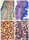
A) Sagittal section of esophagus of Torpedo marmorata Risso embryo at stage I (not flattened, total length 2 cm, external gill filaments). Galgano’s trichromic stain; lymphomyeloid tissue (Lm) appears as a thick band in the submucosa around the esophagus, up to the cranial part of the stomach; the tissue close to the epithelium circumscribes a central lumen; scale bar: 7.8 μm. B) Lymphoidlike cells (arrow) in the area below the lining epithelium (Ep) of the esophagus, star-like mesenchymal cells, close to the external esophageal wall, and several cells in mitosis (arrowhead); scale bar: 6.5 μm. C) Cross section of esophagus of Torpedo marmorata Risso embryo at stage II (flattened embryo, total length 3.2 cm); Galgano’s trichromic stain; lymphoid-like cells located below the lining epithelium of the esophageal canal and mesenchymal cells in the peripheral region are distinguishable; the muscular layer and the external epithelial wall are in the peripheral zone; scale bar: 7.8 μm. D) Mitosis of lymphoid-like cells (arrow); scale bar: 6.5 μm. E) Sagittal section of esophagus of Torpedo marmorata Risso embryo at stage III (flattened, total length 11cm, ready to be born with completely reabsorbed yolk sac); Galgano’s trichromic stain; the lymphomyeloid tissue (Lm) is located between the connective tissue (blue) and the external muscular layer; scale bar: 7.8 μm. F) Neutrophil myelopoiesis (arrow) and eosinophilic myelopoiesis (white arrowhead) are observed; scale bar: 6.5 μm.
Figure 2.
Figure 2.
Immunostaining of consecutive sagittal sections of the external yolk sac of a stage II embryo (flattened embryo, total length 3.2 cm). A) Transverse section of external yolk sac showing the vessels full of megalocytes and area with dispersed yolk drops. Double immunostaining CD34-HRP-DAB/CD38-ALP; scale bar: 10 μm. B) Detail of panel A; CD34+/CD38- cell was detected (arrow); scale bar: 11 μm. C) CD34+/CD38- cell among megalocytes in a vessel (arrow); scale bar: 15 μm.
Figure 3.
Figure 3.
Sagittal sections of epigonal and Leydig organs of adult Torpedo marmorata Risso. A) Lymphomyeloid (Lm) epigonal tissue, surrounding testis, below a part of seminiferous tubule (T) showing spermatozoa; Galgano’s trichromic stain; scale bar: 10 μm. B) Lymphomyeloid (Lm) Leydig tissue, between the connective tissue (blue) and the external muscular layer; Galgano’s trichromic stain; scale bar: 22 μm. C,D) Granulocytopoiesis in epigonal and Leydig lymphomieloid tissues, respectively; scale bar: 6.6 μm.
Figure 4.
Figure 4.
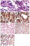
Sagittal sections of esophagus of Torpedo marmorata Risso embryo at stage III (flattened, total length 11cm, ready to be born with completely reabsorbed yolk sac). A) Hemalum-eosin stain. The figure shows cells in various maturation stages, in particular metamyelocytes (white arrowhead) and locus of lymphocytopoiesis (black arrowhead); scale bar: 6.6 μm. B) Myeloid cells at various stages of maturation, in which there are myeloblasts (white arrowhead), myelocytes (white arrow), metamyelocytes (black arrow) and a myeloid cell in mitosis (black arrowhead); no stem cells are detectable with histological staining; scale bar: 6.6 μm. C, D) Double immunostaining CD34-HRP-DAB/CD38-ALP of sections from an embryonic esophagus of stage III; in this section several cell populations can be observed: CD34+/CD38-cells (brown-stained immunoreactivity, black arrowhead), CD34+/CD38+cells (red and brown stained cytoplasm, black arrow), CD34-/CD38+cells (red stained cytoplasm, white arrow), CD34-/CD38-cells (blue, as contrast staining to Hemalum-eosin, white arrowhead); scale bar: 6.6 μm. E) Double immunogold staining CD34-colloidal gold-S.E./CD38-ALP. Four cell phenotypes are observed in this section: CD34+/CD38- (black arrowhead), CD34+/CD38+ (black arrow), CD34-/CD38+ (white arrow) and CD34-/CD38- (white arrowhead). scale bar: 6.6 μm. F) Lymphomyeloid tissue and lymphocytopoiesis; scale bar: 4 μm. G) Three cell phenotypes are observed: CD34+/CD38-cells (black arrowhead), CD34+/CD38+cells (black arrow), CD34-/CD38- cells (white arrowhead); scale bar: 6.6 μm.

References

    1. Zon Li. Developmental biology of hematopoiesis. Blood 1995;86:2876-91. - PubMed
    1. Dzierzak E, Medvinsky A, de Bruijn M. Qualitative and quantitative aspects of haematopoietic cell development in the mammalian embryo. Immunol Today 1998;19:228-36. - PubMed
    1. Tavian M, Coulombel L, Luton D, Clemente HS, Dieterlen-Lievre F, Peault B. Aorta-associated CD34+ hematopoietic cells in the early human embryo. Blood 1996;87:67-72. - PubMed
    1. Tavian M, Hallais MF, Peault B. Emergence of intraembryonic hematopoietic precursors in the preliver human embryo. Development 1999;126:793-803. - PubMed
    1. Barcena A, Muench MO, Kapidzic M, Fisher SJ. A new role for the human placenta as a hematopoietic site throughout gestation. Reprod Sci 2009;16:178-87. doi: 10.1177/1933719108327621. - PMC - PubMed