. 2019 Nov 8;5(11):1963-1974.
doi: 10.1021/acsinfecdis.9b00300. Epub 2019 Oct 2.

Determinants of Active-Site Inhibitor Interaction with HIV-1 RNase H

Affiliations

Determinants of Active-Site Inhibitor Interaction with HIV-1 RNase H

Zhaoyong Xi et al. ACS Infect Dis. .

Abstract

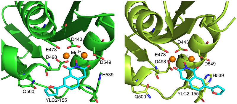
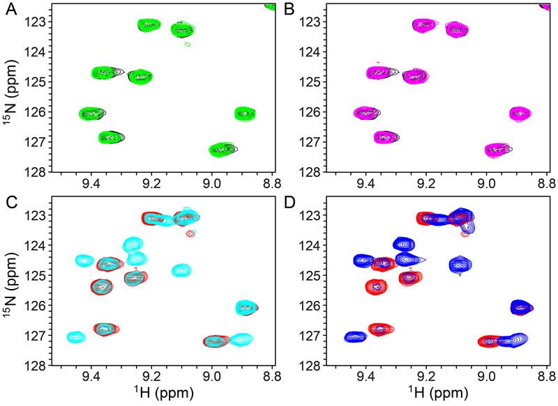
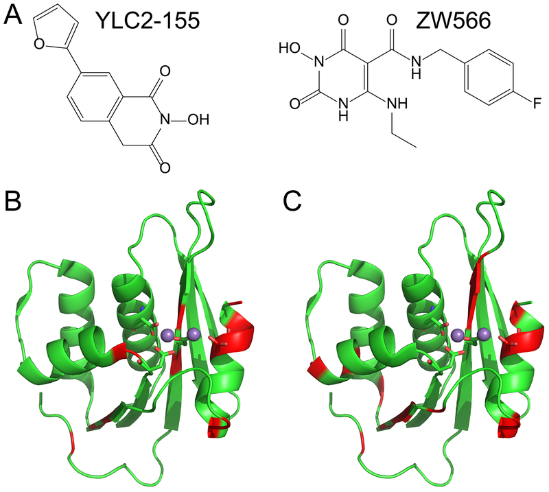
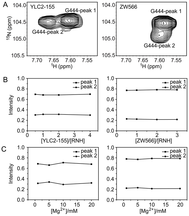
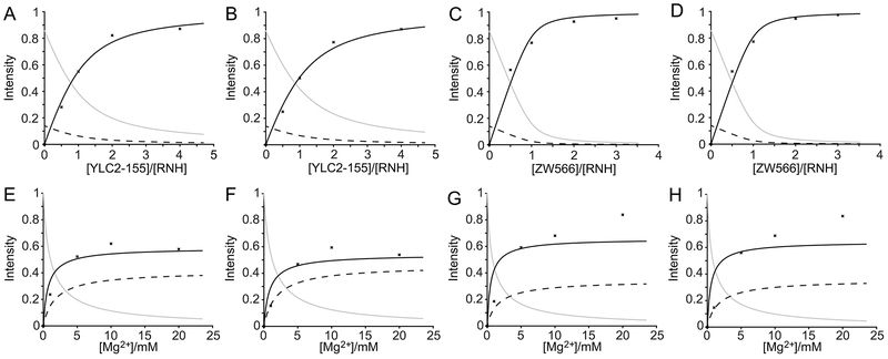
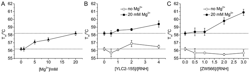
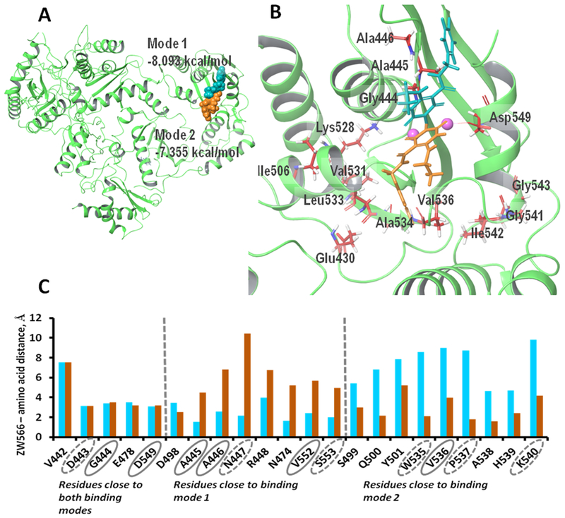
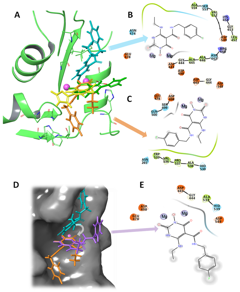
The ribonuclease H (RNH) activity of HIV-1 reverse transcriptase (RT) is essential for viral replication and can be a target for drug development. Yet, no RNH inhibitor to date has substantial antiviral activity to allow advancement into clinical development. Herein, we describe our characterization of the detailed binding mechanisms of RNH active-site inhibitors, YLC2-155 and ZW566, that bind to the RNH domain through divalent metal ions, using NMR, molecular docking, and quantum mechanical calculations. In the presence of Mg2+, NMR spectra of RNH exhibited split (two) resonances for some residues upon inhibitor binding, suggesting two binding modes, an observation consistent with the docking results. The relative populations of the two binding conformers were independent of inhibitor or Mg2+ concentration, with one conformation consistently more favored. In our docking study, one distinctive pose of ZW566 showed more interactions with surrounding residues of RNH compared to the analogous binding pose of YLC2-155. Inhibitor titration experiments revealed a lower dissociation constant for ZW566 compared to YLC2-155, in agreement with its higher inhibitory activity. Mg2+ titration data also indicated a stronger dependence on Mg2+ for the RNH interaction with ZW566 compared to YLC2-155. Combined docking and quantum mechanical calculation results suggest that stronger metal coordination as well as more protein-inhibitor interactions may account for the higher binding affinity of ZW566. These findings support the idea that strategies for the development of potent competitive active site RNH inhibitors should take into account not only metal-inhibitor coordination but also protein-inhibitor interaction and conformational selectivity.

Keywords: HIV; NMR; RNase H; molecular docking; quantum mechanical calculations.


Figures

Figure 1.
Figure 1.
View of the two binding modes of YLC2-155 to the RNH domain, observed in a crystal structure of the complex (5UV5). Mn2+ was used for crystal structure determination.
Figure 2.
Figure 2.
Selected region of 1H-15N HSQC spectra of 50 μM 15N-labeled RNH in the absence (A and B, black) and presence of 20 mM Mg2+ (C and D, red). 50 μM YLC2-155 (green in A and cyan in C) or ZW566 (magenta in B and blue in D) was added. All spectra were recorded at 293 K. Note, we selected a region that does not include split signals in panel C and D to simply explain spectral changes first. See Figure S1 and S2 for overall spectral changes.
Figure 3.
Figure 3.
Structures of the RNH active-site inhibitors used in this study are shown in (A). Residues showing split resonances upon binding of (B) YLC2-155 or (C) ZW566 are mapped onto the crystal structure of the RNH domain (PDB 5UV5), and colored in red. Metal ions are shown as balls, and metal binding residues are displayed in stick representation.
Figure 4.
Figure 4.
Analysis of proportions of two binding modes for residue G444 in the (left column) YLC2-155 bound form and (right column) ZW566 bound form. (A) Peak splitting of residue G444. Normalized peak intensities plotted as a function of (B) [inhibitor]/[RNH] molar ratios or (C) Mg2+ concentration.
Figure 5.
Figure 5.
Inhibitor titration data points and the curve fits for residue F440 (A, C, E and G) and T470 (B, D, F and H) in YLC2-155 (A, B, E and F) or ZW566 (C, D, G and H) titrations. Inhibitor titration was performed in the presence of 50 μM RNH and 20 mM Mg2+. Mg2+ titration was performed in the presence of 50 μM RNH and 50 μM inhibitor. The titration data were analyzed assuming three species: free RNH, RNH-2Mg2+, and RNH-2Mg2+-inhibitor. Normalized peak intensities are plotted as a function of total [inhibitor]/[RNH] molar ratios or Mg2+ concentration. Plots for fitting fractions of RNH, RNH-2Mg2+, and RNH-2Mg2+-Inhibitor are shown as solid gray, dashed black, and solid black line, respectively. Determination of inhibitor dissociation constants (K2) were performed by assuming a two-step mechanism (Equation 1 and 2).
Figure 6.
Figure 6.
Analysis of RNH thermal stability upon Mg2+ and inhibitor binding. Tm values are plotted against (A) Mg2+ concentration, (B) molar ratio of [YLC2-155]/[RNH], and (C) molar ratio of [ZW566]/[RNH].
Figure 7.
Figure 7.
Docking simulations show two binding modes for ZW566. (A) Overview of ZW566 binding in the RNH active site. In binding mode 1 (−8.09 kcal/mol, cyan structure), the fluorinated benzene ring of ZW566 points toward the upper part of p66 subunit. In binding mode 2 (−7.36 kcal/mol, orange structure), the fluorinated benzene ring of ZW566 points toward the p66/p51 interface. (B) A close view of the two ZW566 binding modes with surrounding residues that show split NMR signals. (C) Distances from each of the indicated residues to the nearest proximity of ZW566 in mode 1 (cyan columns) and in mode 2 (orange columns). Residues circled with a solid line show split NMR signals; residues circled with a dashed line are adjacent to residues showing split NMR signals.
Figure 8.
Figure 8.
The two binding modes of ZW566 are distinctive from the two-mode binding modes of YLC2-155. A) Overlap of ZW566 (cyan and orange structures) and YLC2-155 (yellow and green structures) binding modes. B) Ligand interaction diagram (LID) for ZW566 binding mode 1. C) LID for ZW566 binding mode 2. D) ZW566 binding mode 1 (cyan structure), mode 2 (orange structure) and a simulated “YLC-2155-like binding mode” for ZW566 (purple structure) obtain by superposition of the ZW566 chelating ring in binding mode 1 with the ZW566 chelating ring in binding Mode 2. Structures are shown on the RNH active site surface. E) LID for ZW566 superimposed to simulate YLC2-155 binding mode. Such a binding mode would result in fewer interactions with amino acid residues and greater exposure to solvent. In LID, negatively charged residues are shown in red, positively charged residues are shown in blue, hydrophobic residues are shown in green, polar residues are shown in cyan, and glycine is shown in white. Note, in the LID presentation, residue numbers were embedded into the original graphics. In panel C, dashed residue number indicates that from p51 subunit in RT.

Similar articles

Cited by

References

    1. Baltimore D (1970) RNA-dependent DNA polymerase in virions of RNA tumour viruses. Nature 226, 1209–1211. - PubMed
    1. Temin HM, and Mizutani S (1970) RNA-dependent DNA polymerase in virions of Rous sarcoma virus. Nature 226, 1211–1213. - PubMed
    1. Temin HM (1993) Retrovirus variation and reverse transcription: abnormal strand transfers result in retrovirus genetic variation. Proc. Natl. Acad. Sci. U. S. A 90, 6900–6903. - PMC - PubMed
    1. Wilhelm M, and Wilhelm FX (2001) Reverse transcription of retroviruses and LTR retrotransposons. Cell. Mol. Life Sci 58, 1246–1262. - PMC - PubMed
    1. Eickbush TH, and Jamburuthugoda VK (2008) The diversity of retrotransposons and the properties of their reverse transcriptases. Virus Res. 134, 221–234. DOI: 10.1016/j.virusres.2007.12.010 - DOI - PMC - PubMed

Publication types

MeSH terms