. 2019 Dec;33(12):13775-13793.
doi: 10.1096/fj.201901730R. Epub 2019 Oct 2.

AAV2-BDNF promotes respiratory axon plasticity and recovery of diaphragm function following spinal cord injury

Affiliations

AAV2-BDNF promotes respiratory axon plasticity and recovery of diaphragm function following spinal cord injury

Brittany A Charsar et al. FASEB J. 2019 Dec.

Abstract

More than half of spinal cord injury (SCI) cases occur in the cervical region, leading to respiratory dysfunction due to damaged neural circuitry that controls critically important muscles such as the diaphragm. The C3-C5 spinal cord is the location of phrenic motor neurons (PhMNs) that are responsible for diaphragm activation; PhMNs receive bulbospinal excitatory drive predominately from supraspinal neurons of the rostral ventral respiratory group (rVRG). Cervical SCI results in rVRG axon damage, PhMN denervation, and consequent partial-to-complete paralysis of hemidiaphragm. In a rat model of C2 hemisection SCI, we expressed the axon guidance molecule, brain-derived neurotrophic factor (BDNF), selectively at the location of PhMNs (ipsilateral to lesion) to promote directed growth of rVRG axons toward PhMN targets by performing intraspinal injections of adeno-associated virus serotype 2 (AAV2)-BDNF vector. AAV2-BDNF promoted significant functional diaphragm recovery, as assessed by in vivo electromyography. Within the PhMN pool ipsilateral to injury, AAV2-BDNF robustly increased sprouting of both spared contralateral-originating rVRG axons and serotonergic fibers. Furthermore, AAV2-BDNF significantly increased numbers of putative monosynaptic connections between PhMNs and these sprouting rVRG and serotonergic axons. These findings show that targeting circuit plasticity mechanisms involving the enhancement of synaptic inputs from spared axon populations is a powerful strategy for restoring respiratory function post-SCI.-Charsar, B. A., Brinton, M. A., Locke, K., Chen, A. Y., Ghosh, B., Urban, M. W., Komaravolu, S., Krishnamurthy, K., Smit, R., Pasinelli, P., Wright, M. C., Smith, G. M., Lepore, A. C. AAV2-BDNF promotes respiratory axon plasticity and recovery of diaphragm function following spinal cord injury.

Keywords: SCI; cervical; phrenic; sprouting.


Conflict of interest statement

The authors thank Dr. Rich Smeyne (Thomas Jefferson University) for sharing his expertise in 2-dimensional stereology counting techniques, and for his help with implementing these methods, and Dr. Robert Sterling (Thomas Jefferson University) for his patience and assistance with statistical analysis. This work was supported by the U.S. National Institutes of Health, National Institute of Neurological Disorders and Stroke (NINDS; 2R01NS079702 to A.C.L. and 1F30NS103436 to B.A.C.), the Craig H. Neilsen Foundation (476686 to A.C.L.), and the Shriners Hospitals for Children Viral Core Grant SHC 84051 (to G.M.S.). The authors declare no conflicts of interest.

Figures

Figure 1
Figure 1
Intraspinal AAV2-BDNF injection focally enhanced BDNF expression within the PhMN pool. A) Illustration of intraspinal delivery of AAV2 to the ventral horn of spinal cord levels C3–C5 10 d after C2 hemisection. B) GFP reporter expression from AAV2 vector in the ventral horn on the side of injection. Scale bar, 250 μm. CE) Sagittal section of rat spinal cord 8 wk postinjury with GFP reporter and PhMN pool labeled with CTB. E) GFP spread was restricted mostly to the ventral horn. Scale bar, 1 mm. The most rostral point of the PhMN pool was located 2.03 mm (sd ± 0.26 mm, n = 3) from the caudal lesion border. F) Histogram of GFP + area of fluorescence in 500-μm bins throughout the length of the spinal cord in relation to the most rostral point of the PhMN pool (n = 3). G) TrkB receptor expression in rVRG neurons that were retrogradely labeled from the ipsilateral C3–C5 ventral horn with FluroGold (F.Gold). Scale bar, 50 μm. H) Bar graph illustrating BDNF protein concentrations in ipsilateral and contralateral spinal cord halves 2 wk after AAV2-BDNF injection (n = 3). Unpaired Student’s t test; t = 4.351. *P = 0.0121.
Figure 2
Figure 2
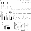
Focal AAV2-BDNF injection promoted recovery of diaphragm function. A) Diaphragmatic EMG recording of ipsilateral hemidiaphragm in intact rats. B, C) Diaphragmatic EMG of ipsilateral hemidiaphragm 8 wk postinjury in AAV2-GFP–treated (B) and AAV2-BDNF–treated (C) rats. Top: raw traces; bottom: integrated signals. D) Quantification of EMG integrated signal for intact (n = 13), injured plus AAV2-GFP (n = 11), and injured plus AAV2-BDNF (n = 10) rats. Repeated measures 2-way ANOVA (post hoc Fisher’s least significant difference test): treatment, F(2,31) = 21.21, P < 0.0001; intact vs. AAV2-GFP, P < 0.0001, t = 6.44; intact vs. AAV2-BDNF, P = 0.0008, t = 3.725; AAV2-GFP vs. AAV2-BDNF, P = 0.0199, t = 2.456; region, F(2,62) = 0.572, P = 0.567.E, F) CMAP recordings of ipsilateral hemidiaphragm after supramaximal phrenic nerve stimulation in AAV2-GFP– treated (E) and AAV2-BDNF–treated (F) rats 8 wk post-SCI (n = 5/group). G) Quantification of CMAP amplitude. Ns, not significant. Unpaired Student’s t test, P = 0.0528, t = 2.271, df = 8. A subset of rats in each injury group was assessed weekly for grip strength, beginning 1 wk prior to injury (baseline = preinjury). H) No difference was found in recovery between AAV2-GFP (n = 3) and AAV2-BDNF–treated (n = 4) C2 hemisection rats. Repeated measures 2-way ANOVA: treatment, F(1,5) = 0.1646, P = 0.702. *P < 0.05; ***P < 0.001; ****P < 0.0001.
Figure 3
Figure 3
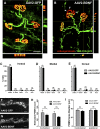
AAV2-BDNF injection did not alter diaphragm NMJ innervation, PhMN survival, or PhMN somal morphology. Synaptic vesicle protein 2 and SMI-312 as markers for presynaptic terminals and neurofilament, respectively, were used to identify the presynaptic component of the diaphragm NMJ. A, B) α-Bungarotoxin was used to label nicotinic acetylcholine receptors to identify postsynaptic receptors of the diaphragm NMJ in hemidiaphragms of AAV2-GFP–treated (n = 4) (A) and AAV2-BDNF–treated (n = 5) (B) rats. Scale bar, 30 μm. Arrowheads (B) denote an example of an NMJ that is partially denervated (arrowheads point to the portion of this NMJ that is denervated: no overlap of green and red labeling). NMJs were analyzed to determine the percentage of intact, partially denervated (P. Denv), and fully denervated (Denv) NMJs, in addition to the percentage of terminal sprouting and multiple axons. CE) Quantification of ipsilateral NMJ morphologies showed no difference in all 3 regions of the hemidiaphragm between AAV2-GFP and AAV2-BDNF rats. Repeated measures 2-way ANOVA: intact, F(1,7) = 0.298, P = 0.602; partially denervated, F(1,7) = 0.269, P = 0.620; fully denervated, F(1,7) = 0.777, P = 0.407; thin preterminal staining (T.S.), F(1,7) = 1.597, P = 0.247; NMJ innervated by multiple axons (M.A.), F(1,7) = 1.263, P = 0.298. F, G) CTB-labeled PhMNs ipsilateral to hemisection in injured rats with AAV2-GFP (F) or AAV2-BDNF (G). H) Quantification of estimated ipsilateral and contralateral PhMN cell counts in AAV2-GFP–treated (n = 8) and AAV2-BDNF–treated (n = 6) rats. Two-way ANOVA: treatment P = 0.2062, F(1,10) = 1.828; side P = 0.523, F(1,10) = 0.4385. I) Quantification of CTB + PhMN soma size. Two-way ANOVA: treatment P = 0.4604, F(1,10) = 0.5893; side P = 0.511, F(1,10) = 0.4642. Ns, not significant.
Figure 4
Figure 4
AAV2-BDNF injection did not promote rVRG axon regeneration. A) Schematic of AAV2-mCherry injection for labeling of ipsilateral-originating rVRG axons. B) Schematic of transverse spinal cord. B′, B″) Representative images of transverse spinal cord 500-μm caudal-to-caudal lesion border demonstrate hemisection completeness. mCherry + puncta can be found in ventral white matter of the contralateral uninjured side of the cervical spinal cord after rVRG labeling with AAV2-mCherry (B′), whereas no mCherry + axons are present on the side of injury (B″). Scale bar, 100 μm. C) mCherry-labeled axons from the ipsilateral rVRG 8 wk postinjury in AAV2-BDNF–treated animal. Scale bar, 500 μm (C, C′). Average lesion width was 430 μm. D) Quantification of number of mCherry + axon profiles in AAV2-GFP (n = 3) and AAV2-BDNF (n = 3) rats. Two-way ANOVA (post hoc Fisher’s least significant difference test); treatment P = 0.4841, F(1,4) = 0.5934. MN, motor neuron.
Figure 5
Figure 5
AAV2-BDNF injection promoted significant sprouting of spared rVRG axons locally within the PhMN pool. A) Schematic of AAV2-mCherry injection for labeling of spared contralateral-originating rVRG axons. B, C) Transverse image of mCherry + axons around CTB + PhMNs in the contralateral cervical spinal cord after injection of AAV2-mCherry into the left rVRG and right C2 hemisection. Scale bar, 50 μm. DE′) Confocal images of mCherry + rVRG axon bundles around denervated CTB + PhMNs (right side) 8 wk after C2 hemisection in AAV2-GFP (D, D′) and AAV2-BDNF (E, E′) rats. Scale bar, 100 μm. F) Quantification of area of mCherry + axons in a 164-μm diameter radius around the CTB PhMNs on the side of hemisection (n = 3/group). Unpaired Student’s t test; P = 0.02, t = 3.654, df = 4. MN, motor neuron. *P < 0.05.
Figure 6
Figure 6
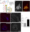
AAV2-BDNF injection significantly enhanced putative excitatory synaptic input to PhMNs from spared rVRG axons. A, B) Representative confocal images with orthogonal projections of CTB/dsRed/VGlut2 triple labeling on the side of hemisection in AAV2-GFP (A) and AAV2-BDNF (B) rats. Scale bar, 20 μm. Yellow arrows indicate colocalization of VGlut2+/mCherry + puncta directly presynaptic to CTB-labeled PhMNs. C) Location of PhMNs in ventral horn for synaptic connection analysis. D) Quantification of number of VGlut2+/mCherry + presynaptic puncta per CTB + PhMN (n = 3 rats/group). Unpaired Student’s t test; P = 0.0043, t = 5.837, df = 4. *P < 0.01.
Figure 7
Figure 7
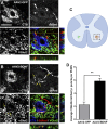
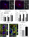
AAV2-BDNF injection significantly enhanced putative synaptic input to PhMNs from sprouting serotonergic axons. A, B) Total area of 5-HT + axons was determined on the side of C2 hemisection using a modified Sholl analysis from the center of PhMN clusters in AAV2-GFP (A) and AAV2-BDNF (B) rats. Scale bars, 200 μm. C, D) Quantification of 5-HT + axon area in the left/contralateral ventral horn (C) and right/ipsilateral ventral horn (D). Right ventral horn repeated measures 2-way ANOVA (post hoc Fisher’s least significant difference test): treatment P = 0.0107, F(1,4) = 20.38, at +200 μm P = 0.0031, at +300 μm P = 0.0005, at +400 μm P = 0.0020; distance P = 0.1514, F(2,8) = 2.412. Left ventral horn repeated measures 2-way ANOVA (post hoc Fisher’s least significant difference test): treatment P = 0.6042, F(1,4) = 0.3156, at +200 μm P = 0.8017; at +300 μm P = 0.4677; at +400 μm P = 0.7718. E, F) Representative confocal images with orthogonal projections of CTB/Syn1/5-HT triple labeling on the side of hemisection in AAV2-GFP (E) and AAV2-BDNF (F) rats. Scale bar, 20 μm. G) Quantification of Syn1+/5-HT + double-labeled puncta onto CTB + PhMNs (n = 3 rats/group). Unpaired Student’s t test; P = 0.0019, t = 7.274, df = 4. Ns, not significant. **P < 0.01; ***P < 0.001.
Figure 8
Figure 8
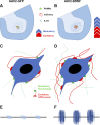
Model of axon plasticity after focal AAV2-BDNF injection. 5-HT axons modulate rhythmic excitatory input onto PhMNs from glutamatergic rVRG axons. A, B) Diagram depicting different forms of input onto PhMNs in AAV2-GFP–treated (A) and AAV2-BDNF–treated (B) rats after a C2 hemisection. C, D) Image of an individual PhMN with rVRG and serotonergic monosynaptic connections. E, F) Effect on diaphragm EMG amplitude after changes in synaptic input to PhMNs.

References

    1. Tator C. H., Fehlings M. G. (1991) Review of the secondary injury theory of acute spinal cord trauma with emphasis on vascular mechanisms. J. Neurosurg. 75, 15–26 - PubMed
    1. Park E., Velumian A. A., Fehlings M. G. (2004) The role of excitotoxicity in secondary mechanisms of spinal cord injury: a review with an emphasis on the implications for white matter degeneration. J. Neurotrauma 21, 754–774 - PubMed
    1. Dobbins E. G., Feldman J. L. (1994) Brainstem network controlling descending drive to phrenic motoneurons in rat. J. Comp. Neurol. 347, 64–86 - PubMed
    1. Smith J. C., Abdala A. P. L., Rybak I. A., Paton J. F. R. (2009) Structural and functional architecture of respiratory networks in the mammalian brainstem. Philos. Trans. R. Soc. Lond. B Biol. Sci. 364, 2577–2587 - PMC - PubMed
    1. Singh A., Tetreault L., Kalsi-Ryan S., Nouri A., Fehlings M. G. (2014) Global prevalence and incidence of traumatic spinal cord injury. Clin. Epidemiol. 6, 309–331 - PMC - PubMed

Publication types

Substances

Supplementary concepts