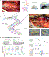
Selectivity of afferent microstimulation at the DRG using epineural and penetrating electrode arrays
- PMID: 31577993
- PMCID: PMC9131467
- DOI: 10.1088/1741-2552/ab4a24
Selectivity of afferent microstimulation at the DRG using epineural and penetrating electrode arrays
Abstract
Objective: We have shown previously that microstimulation of the lumbar dorsal root ganglia (L5-L7 DRG) using penetrating microelectrodes, selectively recruits distal branches of the sciatic and femoral nerves in an acute preparation. However, a variety of challenges limit the clinical translatability of DRG microstimulation via penetrating electrodes. For clinical translation of a DRG somatosensory neural interface, electrodes placed on the epineural surface of the DRG may be a viable path forward. The goal of this study was to evaluate the recruitment properties of epineural electrodes and compare their performance with that of penetrating electrodes. Here, we compare the number of selectively recruited distal nerve branches and the threshold stimulus intensities between penetrating and epineural electrode arrays.
Approach: Antidromically propagating action potentials were recorded from multiple distal branches of the femoral and sciatic nerves in response to epineural stimulation on 11 ganglia in four cats to quantify the selectivity of DRG stimulation. Compound action potentials (CAPs) were recorded using nerve cuff electrodes implanted around up to nine distal branches of the femoral and sciatic nerve trunks. We also tested stimulation selectivity with penetrating microelectrode arrays implanted into ten ganglia in four cats. A binary search was carried out to identify the minimum stimulus intensity that evoked a response at any of the distal cuffs, as well as whether the threshold response selectively occurred in only a single distal nerve branch.
Main results: Stimulation evoked activity in just a single peripheral nerve through 67% of epineural electrodes (35/52) and through 79% of the penetrating microelectrodes (240/308). The recruitment threshold (median = 9.67 nC/phase) and dynamic range of epineural stimulation (median = 1.01 nC/phase) were significantly higher than penetrating stimulation (0.90 nC/phase and 0.36 nC/phase, respectively). However, the pattern of peripheral nerves recruited for each DRG were similar for stimulation through epineural and penetrating electrodes.
Significance: Despite higher recruitment thresholds, epineural stimulation provides comparable selectivity and superior dynamic range to penetrating electrodes. These results suggest that it may be possible to achieve a highly selective neural interface with the DRG without penetrating the epineurium.
Conflict of interest statement
Disclosures
No conflicts of interest, financial or otherwise, are declared by the authors.
Figures


Similar articles
-
Microstimulation of the lumbar DRG recruits primary afferent neurons in localized regions of lower limb.J Neurophysiol. 2016 Jul 1;116(1):51-60. doi: 10.1152/jn.00961.2015. Epub 2016 Apr 6. J Neurophysiol. 2016. PMID: 27052583 Free PMC article.
-
Microstimulation of primary afferent neurons in the L7 dorsal root ganglia using multielectrode arrays in anesthetized cats: thresholds and recruitment properties.J Neural Eng. 2009 Oct;6(5):055009. doi: 10.1088/1741-2560/6/5/055009. Epub 2009 Sep 1. J Neural Eng. 2009. PMID: 19721181 Free PMC article.
-
Chronic recruitment of primary afferent neurons by microstimulation in the feline dorsal root ganglia.J Neural Eng. 2014 Jun;11(3):036007. doi: 10.1088/1741-2560/11/3/036007. Epub 2014 Apr 24. J Neural Eng. 2014. PMID: 24762981
-
Selective electrical interfaces with the nervous system.Annu Rev Biomed Eng. 2002;4:407-52. doi: 10.1146/annurev.bioeng.4.020702.153427. Epub 2002 Mar 22. Annu Rev Biomed Eng. 2002. PMID: 12117764 Review.
-
Neural stimulation and recording electrodes.Annu Rev Biomed Eng. 2008;10:275-309. doi: 10.1146/annurev.bioeng.10.061807.160518. Annu Rev Biomed Eng. 2008. PMID: 18429704 Review.
Cited by
-
Neuromorphic hardware for somatosensory neuroprostheses.Nat Commun. 2024 Jan 16;15(1):556. doi: 10.1038/s41467-024-44723-3. Nat Commun. 2024. PMID: 38228580 Free PMC article. Review.
-
Computational modeling of dorsal root ganglion stimulation using an Injectrode.J Neural Eng. 2024 Apr 11;21(2):026039. doi: 10.1088/1741-2552/ad357f. J Neural Eng. 2024. PMID: 38502956 Free PMC article.
-
Dorsal Root Ganglion Stimulation for Chronic Pain: Hypothesized Mechanisms of Action.J Pain. 2022 Feb;23(2):196-211. doi: 10.1016/j.jpain.2021.07.008. Epub 2021 Aug 20. J Pain. 2022. PMID: 34425252 Free PMC article. Review.
-
High-density spinal cord stimulation selectively activates lower urinary tract nerves.J Neural Eng. 2022 Nov 22;19(6):066014. doi: 10.1088/1741-2552/aca0c2. J Neural Eng. 2022. PMID: 36343359 Free PMC article.
-
Recruitment of Primary Afferents by Dorsal Root Ganglion Stimulation using the Injectrode.Int IEEE EMBS Conf Neural Eng. 2021 May;2021:609-612. doi: 10.1109/ner49283.2021.9441420. Epub 2021 Jun 2. Int IEEE EMBS Conf Neural Eng. 2021. PMID: 34630868 Free PMC article.
References
-
- Ziegler-Graham K, MacKenzie E, Ephraim P, Travison T and Brookmeyer R 2008. Estimating the prevalence of limb loss in the United States: 2005 to 2050 Arch. Phys. Med. Rehabil 89 422–9 - PubMed
-
- Miller WC, Deathe AB, Speechley M and Koval J 2001. The influence of falling, fear of falling, and balance confidence on prosthetic mobility and social activity among individuals with a lower extremity amputation Arch. Phys. Med. Rehabil 82 1238–44 - PubMed
-
- Biddiss E, Beaton D and Chau T 2007. Consumer design priorities for upper limb prosthetics Disabil. Rehabil. Assist. Technol 2 346–57 - PubMed
-
- Cole MJ, Durham S and Ewins D 2008. An evaluation of patient perceptions to the value of the gait laboratory as part of the rehabilitation of primary lower limb amputees Prosthet. Orthot. Int 32 12–22 - PubMed
-
- Raspopovic S. et al. Restoring natural sensory feedback in real-time bidirectional hand prostheses. Sci. Transl. Med. 2014;6:222ra19. - PubMed
Publication types
MeSH terms
Grants and funding
LinkOut - more resources
Full Text Sources
Miscellaneous
