. 2019 Dec 20;294(51):19451-19464.
doi: 10.1074/jbc.RA119.010060. Epub 2019 Oct 2.

JARID2 and the PRC2 complex regulate the cell cycle in skeletal muscle

Affiliations

JARID2 and the PRC2 complex regulate the cell cycle in skeletal muscle

Abhinav Adhikari et al. J Biol Chem. .

Abstract

JARID2 is a noncatalytic member of the polycomb repressive complex 2 (PRC2) which methylates of histone 3 lysine 27 (H3K27). In this work, we show that JARID2 and the PRC2 complex regulate the cell cycle in skeletal muscle cells to control proliferation and mitotic exit. We found that the stable depletion of JARID2 leads to increased proliferation and cell accumulation in S phase. The regulation of the cell cycle by JARID2 is mediated by direct repression of both cyclin D1 and cyclin E1, both of which are targets of PRC2-mediated H3K27 methylation. Intriguingly, we also find that the retinoblastoma protein (RB1) is a direct target of JARID2 and the PRC2 complex. The depletion of JARID2 is not sufficient to activate RB1. However, the ectopic expression of RB1 can suppress cyclin D1 expression in JARID2-depleted cells. Transient depletion of JARID2 in skeletal muscle cells leads to a transient up-regulation of cyclin D1 that is quickly suppressed with no resulting effect on proliferation, Taken together, we show that JARID2 and the PRC2 complex regulate skeletal muscle proliferation in a precise manner that involves the repression of cyclin D1, thus restraining proliferation and repressing RB1, which is required for mitotic exit and terminal differentiation.

Keywords: EZH2; JARID2; Jumonji; cell cycle; chromatin state; cyclin D1; cyclin E1; gene regulation; histone demethylase; polycomb; polycomb repressive complex 2 (PRC2); retinoblastoma protein (pRb, RB); skeletal muscle.


Conflict of interest statement

The authors declare that they have no conflicts of interest with the contents of this article

Figures

Figure 1.
Figure 1.
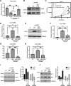
JARID2 depletion results in increased proliferation and DNA synthesis in C2C12 cells. A, an equal number of scrambled (scr) control or short-hairpin RNA against Jarid2 (shJarid2) expressing C2C12 cells were assayed for proliferation on the indicated days. B, scr and shJarid2 C2C12 cells were immunostained for EdU (top panel). Percent of EdU+ nuclei were counted in at least five random fields and plotted (bottom panel). DAPI was used to stain nuclei. Scale bar, 100 μm; n = 3 biological replicates. C, loss of JARID2 increases the G1/S transition during the cell cycle. Cells in A were ethanol fixed, propidium iodide stained, and analyzed for cell cycle phase distribution using flow cytometry. Error bars, mean ± S.E. (Student's t test; ns represents not significant; *, p < 0.05; **, p < 0.01; and ***, p < 0.001; n = 4 biological replicates.)
Figure 2.
Figure 2.
JARID2 inhibits pro-proliferative cell cycle regulators. A and B, JARID2 represses cyclin D1. Scrambled (scr) and shRNA expressing (shJarid2) C2C12 cells were assayed for cyclin D1 by qRT-PCR (A) and Western blotting (B, left panel). B, right panel, blots were quantified and plotted. Plots were normalized to GAPDH loading control. C and D, JARID2 also represses cyclin E1. As in A, cells were assayed for cyclin E1 by qRT-PCR (C) and Western blotting (D, left panel). D, right panel, blots in C were quantified and plotted. Plots were normalized to GAPDH loading control. E–G, JARID2 also represses cyclin-dependent kinases. Cells in A were assayed for Cdk2 and Cdk4 by qRT-PCR (E and G) and Western blotting (F, left panel). D and F were performed simultaneously and the same GAPDH blot was used. Blots in F were quantified and plotted. Plots were normalized to GAPDH (F, right panel). Error bars, mean ± S.E. (Student t test; *, p < 0.05 and ***, p < 0.001; n = 3–6 (qRT-PCR) and 3 (Western blots) biological replicates.)
Figure 3.
Figure 3.
Stable loss of JARID2 down-regulates negative cell cycle regulators in C2C12 cells. A and B, RB1 is inhibited in JARID2-depleted cells. Proliferating scrambled (scr) and JARID2-depleted (shJarid2) C2C12 cells were assayed by qRT-PCR (A) and Western blotting assays (B, left panel). B, right panel, blots were quantified and plotted. Plots were normalized to GAPDH loading control. C and D, Cdkn1A (p21) is repressed in JARID2-depleted cells. Cells in A were assayed for p21 by qRT-PCR (C) and Western blotting (D, left panel). C, right panel, blots in C were quantified and plotted. Plots were normalized to TUBULIN loading control. Error bars, mean ± S.E. (Student's t test; ***, p < 0.001; n = 5 (qRT-PCR) and 3 (Western blots) biological replicates.)
Figure 4.
Figure 4.
JARID2 rescues cell cycle regulator de-repression in JARID2-depleted cells. A and B, Scr and shJarid2 C2C12 cells were transfected with empty vector (pEF) or a Jarid2 (mRNA) expression construct (pEF Jarid2). Cells were assayed by qRT-PCR (A) and Western blotting (B) for Jarid2 expression. TUBULIN was used as the loading control. C, restoration of JARID2 expression rescues proliferation impairment. An equal number of cells in A were assayed for proliferation on indicated time points. D and E, JARID2 rescues cyclin D1 de-repression. Cells in A were assayed for cyclin D1 by qRT-PCR (D) and Western blotting assays (E). TUBULIN was used as the loading control. F, JARID2 rescues cyclin E1 de-repression. Cells in A were assayed for cyclin E1 by qRT-PCR. G and H, cells in A were assayed for Cdk4 (G) and Cdk2 (H) by qRT-PCR. I, Western blot analysis of CDK4 and CDK2 in cells on the restoration of JARID2 expression as in A. TUBULIN was used as the loading control. The blots were quantified and plotted after normalizing to TUBULIN loading control (right panel). J, cells in A were assayed for negative cell cycle regulators, p21 and RB1, by Western blot analysis. TUBULIN was used as a loading control. The blots were quantified and plotted after normalizing to TUBULIN loading control (right panel). Error bars, mean ± S.E. (Analysis of variance test followed by Tukey's multiple comparisons test; ns represents not significant; *, p < 0.05; **, p < 0.01; and ***, p < 0.001, n = 3 biological replicates.)
Figure 5.
Figure 5.
JARID2 directly regulates the expression of cyclin D1 and cyclin E1 via PRC2 recruitment. A–D, ChIP assays were performed on proliferating scr and JARID2-depleted (shJarid2) C2C12 cells with antibodies against JARID2 (A), H3K9me (B), H3K27me3 and H3 (C), EZH2 (D), and primers spanning the Ccnd1 promoter. E, cells in A were assayed for H3K27me3 and H3 status on a HoxB7 region as a positive control. F, cells in A were assayed for the JARID2, EZH2, H3K27me3, and H3K9me status on Tnni2 proximal promoter as a negative control. G, β-catenin recruitment is not enhanced in JARID2-depleted cells. ChIP assay on cells as in A with antibodies against β-catenin and primers spanning the Ccnd1 promoter. H–K, JARID2 also directly regulates cyclin E1 (Ccne1) through PRC2 recruitment. ChIP assays were performed on cells as in A with antibodies against JARID2 (H), H3K9me (I), H3K27me3 and H3 (J), EZH2 (K), and primers spanning the Ccne1 promoter. IgG background signals were subtracted from IP signals. Error bars, mean ± S.E. (Student's t test; ns represents not significant; *, p < 0.05; **, p < 0.01; and ***, p value < 0.001; n = 3–4 biological replicates.)
Figure 6.
Figure 6.
Rb1 is directly regulated by JARID2 but not Cdkn1a (p21). A–D, RB1 is a direct target of JARID2 and the PRC2 complex. ChIP assays were performed on proliferating scr and JARID2-depleted (shJarid2) C2C12 cells with antibodies against JARID2 (A), H3K9me (B), H3K27me3 and H3 (C) EZH2 (D), and primers spanning the Rb1 promoter. IgG background signals were subtracted from immunoprecipitation signals. E, restoration of RB1 can inhibit cyclin D1, but not activate myogenin. C2C12 cells with scrambled (scr) and JARID2 depletion (shJarid2) were transiently transfected with expression constructs for RB1 (RB1-WT), and RB1 mutated at phosphorylation sites (RB1-NPC). UD represents the proliferative, undifferentiated time point and D2′ represents 2 days in differentiation media. Cells were analyzed for total RB1, cyclin D1, and myogenin. GAPDH was used as a loading control. # represents the band of interest for RB1 protein as determined by molecular weight standards. Error bars, mean ± S.E. (Student's t test; ns represents not significant, *, p value < 0.05; **, p value < 0.01; ***, p value < 0.001; n = 3 biological replicates.)
Figure 7.
Figure 7.
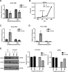
Transient loss of Jarid2 does not decrease cell viability in C2C12 cells. A, C2C12 cells were transiently transfected with scrambled (scr) or shRNA against Jarid2 (shJarid2) without drug selection and were analyzed for JARID2 expression by qRT-PCR at indicated time point post transfection. B, cells in A, were assayed for proliferation rate. C, JARID2 regulates myogenin in C2C12 cells. Cells in A were assayed for myogenin expression by qRT-PCR. D and E, cyclin D1 is regulated by JARID2. D, cells in A were assayed for cyclin D1 expression by qRT-PCR. E, left panel, cells in A were assayed for CYCLIN D1 and JARID2 expression by Western blotting. GAPDH was used as a loading control. E, right panel, blots were quantified and normalized to GAPDH and plotted. Error bars, mean ± S.E. (n = 3 biological replicates.)
Figure 8.
Figure 8.
Transient loss of Jarid2 does not decrease cell viability in primary myoblasts. A, freshly isolated primary myoblasts were transiently transfected with scrambled (scr) control or shRNA against Jarid2 (shJarid2) without drug selection. Jarid2 expression was assayed by qRT-PCR at the indicated time points. B, cells in A were assayed for cyclin D1 expression by qRT-PCR. C, transient loss of Jarid2 in primary myoblast does not have significant change cell cycle progression. Cells in A were ethanol fixed, propidium iodide stained, and analyzed for cell cycle phase distribution using flow cytometry. D, transient loss of Jarid2 did not result in a significant increase in DNA synthesis in primary myoblasts. Cells in A were assayed for DNA synthesis by EdU incorporation assay (left panel). Five random fields were counted for EdU+ nuclei and plotted (right panel). DAPI was used to stain nuclei. Scale bar, 50 μm; n = 2 biological replicates. E–J, cell cycle genes are deregulated upon transient loss of Jarid2 in primary myoblasts. Cells as in A were analyzed for the expression of cyclin E1 (Ccne1) (E), Cdk4 (F), Cdk2 (G), p21 (Cdkn1a) (I), and Cdkn2a (p16/19) (J) by qRT-PCR and Western blotting (H). GAPDH was used as a loading control. # represents the band of interest for CDK2 based on molecular weight standards. K and L, Jarid2 depletion increased apoptotic marker expression in primary myoblasts. Cells in A were assayed for the pro-apoptotic marker Bax (K) and anti-apoptotic marker Bcl2 (L) by qRT-PCR. Error bars, mean ± S.E. (Student's t test; ns represents not significant; ***, p < 0.001; n = 3 biological replicates.)

References

    1. Shen X., Collier J. M., Hlaing M., Zhang L., Delshad E. H., Bristow J., and Bernstein H. S. (2003) Genome-wide examination of myoblast cell cycle withdrawal during differentiation. Dev. Dyn. 226, 128–138 10.1002/dvdy.10200 - DOI - PubMed
    1. Geng Y., Yu Q., Sicinska E., Das M., Bronson R. T., and Sicinski P. (2001) Deletion of the p27Kip1 gene restores normal development in cyclin D1-deficient mice. Proc. Natl. Acad. Sci. U.S.A. 98, 194–199 10.1073/pnas.98.1.194 - DOI - PMC - PubMed
    1. Hitomi M., and Stacey D. W. (1999) Cyclin D1 production in cycling cells depends on ras in a cell-cycle-specific manner. Curr. Biol. 9, 1075–1084 10.1016/S0960-9822(99)80476-X - DOI - PubMed
    1. Berkes C. A., and Tapscott S. J. (2005) MyoD and the transcriptional control of myogenesis. Semin. Cell Dev. Biol. 16, 585–595 10.1016/j.semcdb.2005.07.006 - DOI - PubMed
    1. Lassar A. B., Skapek S. X., and Novitch B. (1994) Regulatory mechanisms that coordinate skeletal muscle differentiation and cell cycle withdrawal. Curr. Opin. Cell Biol. 6, 788–794 10.1016/0955-0674(94)90046-9 - DOI - PubMed

Publication types