

Ventricular volume expansion in presymptomatic genetic frontotemporal dementia
- PMID: 31578297
- PMCID: PMC6946476
- DOI: 10.1212/WNL.0000000000008386
Ventricular volume expansion in presymptomatic genetic frontotemporal dementia
Abstract
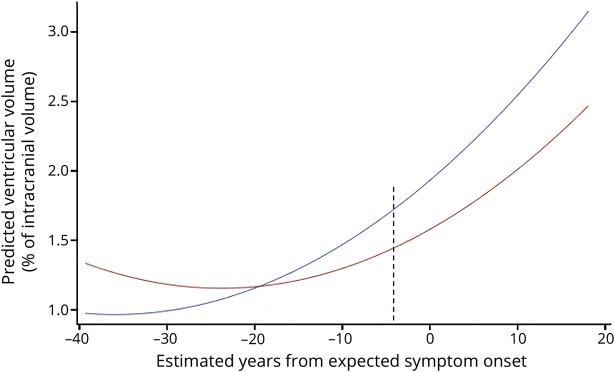
Objective: To characterize the time course of ventricular volume expansion in genetic frontotemporal dementia (FTD) and identify the onset time and rates of ventricular expansion in presymptomatic FTD mutation carriers.
Methods: Participants included patients with a mutation in MAPT, PGRN, or C9orf72, or first-degree relatives of mutation carriers from the GENFI study with MRI scans at study baseline and at 1 year follow-up. Ventricular volumes were obtained from MRI scans using FreeSurfer, with manual editing of segmentation and comparison to fully automated segmentation to establish reliability. Linear mixed models were used to identify differences in ventricular volume and in expansion rates as a function of time to expected disease onset between presymptomatic carriers and noncarriers.
Results: A total of 123 participants met the inclusion criteria and were included in the analysis (18 symptomatic carriers, 46 presymptomatic mutation carriers, and 56 noncarriers). Ventricular volume differences were observed 4 years prior to symptom disease onset for presymptomatic carriers compared to noncarriers. Annualized rates of ventricular volume expansion were greater in presymptomatic carriers relative to noncarriers. Importantly, time-intensive manually edited and fully automated ventricular volume resulted in similar findings.
Conclusions: Ventricular volume differences are detectable in presymptomatic genetic FTD. Concordance of results from time-intensive manual editing and fully automatic segmentation approaches support its value as a measure of disease onset and progression in future studies in both presymptomatic and symptomatic genetic FTD.
Copyright © 2019 The Author(s). Published by Wolters Kluwer Health, Inc. on behalf of the American Academy of Neurology.
Figures
References
-
- Rohrer JD. Structural brain imaging in frontotemporal dementia. Biochim Biophys Acta 2012;1822:325–332. - PubMed