. 2019 Oct 2;9(1):14168.
doi: 10.1038/s41598-019-50793-x.

Widespread brain reorganization perturbs visuomotor coordination in early glaucoma

Affiliations

Widespread brain reorganization perturbs visuomotor coordination in early glaucoma

Vivek Trivedi et al. Sci Rep. .

Abstract

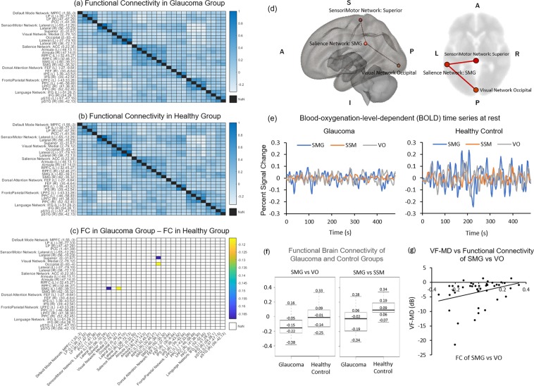
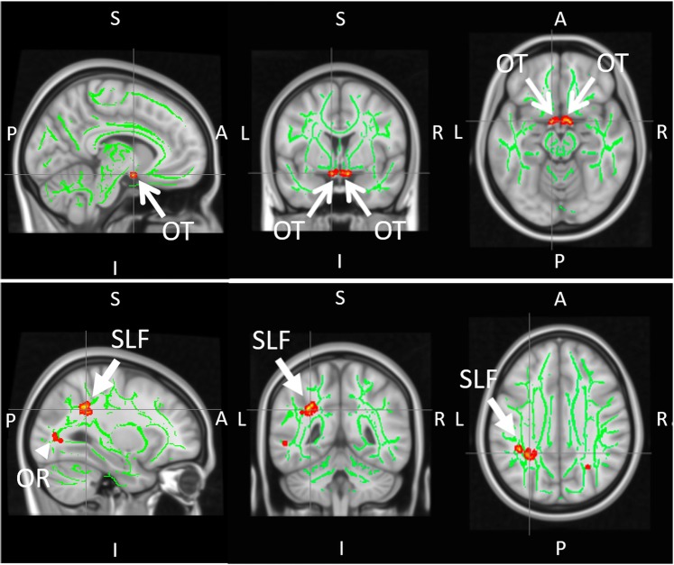
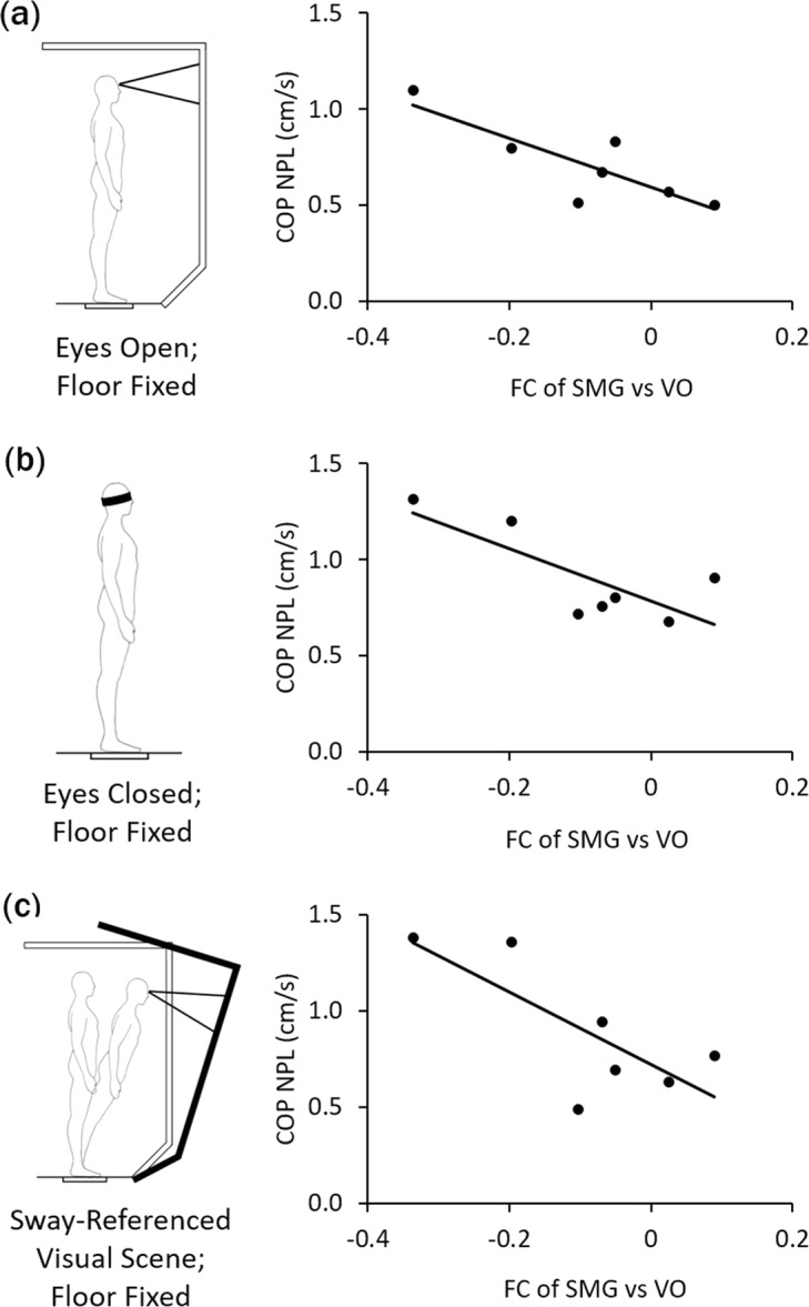
Glaucoma is the world's leading cause of irreversible blindness, and falls are a major public health concern in glaucoma patients. Although recent evidence suggests the involvements of the brain toward advanced glaucoma stages, the early brain changes and their clinical and behavioral consequences remain poorly described. This study aims to determine how glaucoma may impair the brain structurally and functionally within and beyond the visual pathway in the early stages, and whether these changes can explain visuomotor impairments in glaucoma. Using multi-parametric magnetic resonance imaging, glaucoma patients presented compromised white matter integrity along the central visual pathway and around the supramarginal gyrus, as well as reduced functional connectivity between the supramarginal gyrus and the visual occipital and superior sensorimotor areas when compared to healthy controls. Furthermore, decreased functional connectivity between the supramarginal gyrus and the visual brain network may negatively impact postural control measured with dynamic posturography in glaucoma patients. Taken together, this study demonstrates that widespread structural and functional brain reorganization is taking place in areas associated with visuomotor coordination in early glaucoma. These results implicate an important central mechanism by which glaucoma patients may be susceptible to visual impairments and increased risk of falls.


Conflict of interest statement

J.S.S. has made the following disclosure: Royalties e Zeiss, Dublin, CA (for intellectual property licensed by the Massachusetts Institute of Technology and Massachusetts Eye and Ear Infirmary).

Figures

Figure 1
Figure 1
Functional brain connectivity in glaucoma (n = 32) and healthy control (n = 10) subjects. (a–c) Heat maps showing correlation coefficients (r) between temporal profiles for each pair of brain regions-of-interest (ROIs) in glaucoma subjects (a), healthy controls (b), and the difference between glaucoma subjects and healthy control (glaucoma – control) (c) during task-free functional MRI. The coordinates in brackets indicate the center of ROIs in Montreal Neurological Institute (MNI) space. Details of the seed and target brain regions can be found in Supplementary Information 2. In (c), correlation coefficients are shown only for ROI pairs with significant group differences; (d) Three-dimensional sagittal (left) and 2-dimensional axial (right) views of the brain showing the locations of the left supramarginal gyrus (SMG), the visual occipital area (VO), and the superior sensorimotor area (SSM); (e) Time series of blood-oxygenation-level-dependent (BOLD) signal percent change for SMG (blue), SSM (orange), and VO (grey) in glaucoma subjects (left) and healthy controls (right); (f) Box and whisker plots showing functional connectivity (FC) between SMG and VO areas (left) or between SMG and SSM areas (right) in terms of correlation coefficients (r) for both glaucoma and healthy control subjects. In descending order, values represent: maximum, third quartile, median, first quartile, and minimum. FC between SMG and SSM was greater than zero in healthy controls (p < 0.05) but was not different from zero for glaucoma subjects (p = 0.07). Conversely, FC between SMG and VO was less than zero in glaucoma subjects (p < 0.001) but was not different from zero in healthy controls (p = 0.48); (g) Scatter plot showing association between visual field mean deviation (VF-MD) and FC between SMG and VO areas for all subjects.
Figure 2
Figure 2
Group comparisons of white matter integrity between glaucoma patients (n = 32) and healthy controls (n = 10) using tract-based spatial statistics (TBSS) of fractional anisotropy (FA) maps in diffusion tensor MRI. Green pixels represent the FA skeletons of major tracts overlaid on the anatomical brain images in grayscale in the sagittal (left column), coronal (middle column) and axial planes (right column). Red/yellow pixels indicate white matter tract regions that had lower FA in glaucoma patients compared to healthy controls (p < 0.05). These regions include the central visual pathway in the optic tracts (OT; open arrows) and optic radiation (OR; arrowhead) (top row), as well as the left superior longitudinal fasciculus (SLF; closed arrows) around the supramarginal gyrus in the non-visual pathway (bottom row). (A: anterior; P: posterior; L: left; R: right; S: superior; I: inferior).
Figure 3
Figure 3
Postural control in glaucoma subjects (n = 7). Scatter plots showing the brain-behavior associations between functional connectivity (FC) of supramarginal gyrus (SMG) versus visual occipital (VO) areas and sway velocity [i.e. normalized path length (NPL) of the center of pressure (COP)] during dynamic posturography under three conditions: (a) fixed floor and eyes open (r = −0.83, p < 0.05); (b) fixed floor and eyes closed (r = −0.77, p < 0.05); and (c) fixed floor in sway-referenced visual environment with altering visual information (r = −0.75, p = 0.05).

References

    1. Tham Y-C, et al. Global prevalence of glaucoma and projections of glaucoma burden through 2040: a systematic review and meta-analysis. Ophthalmology. 2014;121:2081–2090. doi: 10.1016/j.ophtha.2014.05.013. - DOI - PubMed
    1. Susanna R, Jr., De Moraes CG, Cioffi GA, Ritch R. Why Do People (Still) Go Blind from Glaucoma? Transl Vis Sci Technol. 2015;4:1. doi: 10.1167/tvst.4.2.1. - DOI - PMC - PubMed
    1. Faiq, M. A., Wollstein, G., Schuman, J. S. & Chan, K. C. Cholinergic nervous system and glaucoma: From basic science to clinical applications. Prog Retin Eye Res (2019). - PMC - PubMed
    1. Sponsel WE, Groth SL, Satsangi N, Maddess T, Reilly MA. Refined Data Analysis Provides Clinical Evidence for Central Nervous System Control of Chronic Glaucomatous Neurodegeneration. Transl Vis Sci Technol. 2014;3:1. doi: 10.1167/tvst.3.3.1. - DOI - PMC - PubMed
    1. Calkins DJ, Horner PJ. The cell and molecular biology of glaucoma: axonopathy and the brain. Invest Ophthalmol Vis Sci. 2012;53:2482–2484. doi: 10.1167/iovs.12-9483i. - DOI - PMC - PubMed

Publication types