Review
. 2020 Apr;17(4):216-228.
doi: 10.1038/s41569-019-0265-3. Epub 2019 Oct 2.

Transcriptional and epigenetic regulation of macrophages in atherosclerosis

Affiliations
Review

Transcriptional and epigenetic regulation of macrophages in atherosclerosis

Tatyana Kuznetsova et al. Nat Rev Cardiol. 2020 Apr.

Abstract

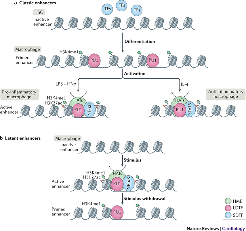
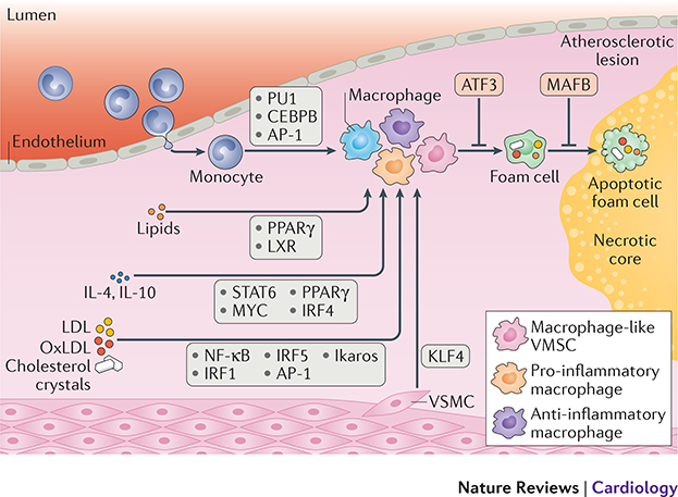
Monocytes and macrophages provide defence against pathogens and danger signals. These cells respond to stimulation in a fast and stimulus-specific manner by utilizing complex cascaded activation by lineage-determining and signal-dependent transcription factors. The complexity of the functional response is determined by interactions between triggered transcription factors and depends on the microenvironment and interdependent signalling cascades. Dysregulation of macrophage phenotypes is a major driver of various diseases such as atherosclerosis, rheumatoid arthritis and type 2 diabetes mellitus. Furthermore, exposure of monocytes, which are macrophage precursor cells, to certain stimuli can lead to a hypo-inflammatory tolerized phenotype or a hyper-inflammatory trained phenotype in a macrophage. In atherosclerosis, macrophages and monocytes are exposed to inflammatory cytokines, oxidized lipids, cholesterol crystals and other factors. All these stimuli induce not only a specific transcriptional response but also interact extensively, leading to transcriptional and epigenetic heterogeneity of macrophages in atherosclerotic plaques. Targeting the epigenetic landscape of plaque macrophages can be a powerful therapeutic tool to modulate pro-atherogenic phenotypes and reduce the rate of plaque formation. In this Review, we discuss the emerging role of transcription factors and epigenetic remodelling in macrophages in the context of atherosclerosis and inflammation, and provide a comprehensive overview of epigenetic enzymes and transcription factors that are involved in macrophage activation.


Conflict of interest statement

Competing interests

None

Figures

Figure 1:
Figure 1:. Epigenetic landscape of regulatory DNA is set-up by transcription factors.
Chromatin is generally divided into transcription permissive euchromatin and compact inactive heterochromatin. Heterochromatin is often associated with repressive histone modifications such as H3K9me2/3 and H3K27me3. Enhancers and promoters are located in euchromatin and are associated with a distinct epigenetic mark-up (Box 2). Top left: active enhancers can be characterized by the presence of broad H3K4me1 and narrow H3K27ac marks and an ATAC-seq (open chromatin) signal. Top right: active promoters display narrow H3K4me3, H3K27Ac, and ATAC signal. Bottom: transcription factor (TF) binding sites are located within the nucleosome-free regions, characterized by ATAC-seq signal enrichment. In macrophages regulatory elements are associated with binding of lineage determining TFs PU1, CEBPB and AP-1 and can be further activated by binding of signal dependent TFs such as LPS-induced NF-κB or lipid-induced PPAR-ɣ. Activating TFs can recruit histone modifying enzymes (HMEs), such as histone acetyl transferases (HATs) to set up a permissive chromatin landscape. At promoters SDTFs do not regularly bind directly, but the open chromatin allows for the binding of RNA polymerase II, poising the gene for transcription.
Figure 2:
Figure 2:. Macrophage enhancer selection. Classic enhancers:
are opened by lineage determining transcription factor (LDTF) PU1 during myeloid lineage differentiation to macrophages. Enhancers are primed by deposition of H3K4me1 by histone modifying enzymes (HMEs) from the lysine methyl transferase (KMT) families 2, 3 or 7 (Box 2). Upon activation, signal dependent transcription factor (SDTFs) such as NF-κB for pro-inflammatory M1-like activation or STAT6 for anti-inflammatory M2-like activation, are recruited to the primed enhancers. This in turn leads to recruitment of histone acetyl transferases (HATs) to set up a permissive chromatin landscape and activate the enhancer. Latent enhancers: are unbound by LDTFs such as PU1 in a steady-state macrophage. Upon stimulus, SDTFs such as NF-κB can bind to their partially uncovered motifs and open up the chromatin, leading to PU1, KMT, and HAT recruitment to fully open up the chromatin and activate the enhancer. After removal of the stimulus, the SDTF leaves the chromatin, and activating histone marks are removed by recruitment of histone de-acetylases (HDACs), however H3K4me1 remains unremoved and keeps enhancer in a primed state for subsequent activation. This enhancer memory enables a quicker activation upon the next encounter of the same stimulus.
Figure 3:
Figure 3:. Transcription factors modulate macrophages in atherosclerosis.
Upon recruitment to an atherosclerotic plaque, circulating monocytes are differentiated into macrophages. Lineage determining transcription factors (LDTFs) such as PU1, CEBPB, and AP-1 mark macrophage specific regulatory elements. Smooth muscle cells migrating into the plaque can also differentiate into a macrophage like state by induction of KLF4 TF. Macrophages can subsequently be activated into a plethora of different phenotypes. Lipid accumulation leads to foam cell formation and upregulation of PPAR-ɣ and LXR. Stimulation with anti-inflammatory triggers such as IL4 and IL10 leads to expression of i.a. STAT6, MYC, PPAR-ɣ, and IRF4. Stimulation by pro-inflammatory triggers such as LPS, oxLDL, and cholesterol leads to activation of i.a. NF-κB, IRFs and IKAROS. Foam cell formation can be inhibited by expression of ATF3 while foam cell apoptosis is inhibited by MAFB.

References

    1. Woollard KJ, Geissmann F. Monocytes in atherosclerosis: subsets and functions. Nat Rev Cardiol. Nature Publishing Group; 2010. February;7(2):77–86. - PMC - PubMed
    1. Gordon S, Plüddemann A. Tissue macrophages: heterogeneity and functions. BMC Biology; 2017. June 22;:1–18. - PMC - PubMed
    1. Ginhoux F, Schultze JL, Murray PJ, Ochando J, Biswas SK. New insights into the multidimensional concept of macrophage ontogeny, activation and function. Nat Immunol. Nature Publishing Group; 2016. January;17(1):34–40. - PubMed
    1. Xue J, Schmidt SV, Sander J, Draffehn A, Krebs W, Quester I, De Nardo D, Gohel TD, Emde M, Schmidleithner L, Ganesan H, Nino-Castro A, Mallmann MR, Labzin L, Theis H, Kraut M, Beyer M, Latz E, Freeman TC, Ulas T, Schultze JL. Transcriptome-Based Network Analysis Reveals a Spectrum Model of Human Macrophage Activation. Immunity. Elsevier Inc; 2014. February 20;40(2):274–88. - PMC - PubMed
    1. Colin S, Chinetti-Gbaguidi G, Staels B. Macrophage phenotypes in atherosclerosis. Immunol Rev. John Wiley & Sons, Ltd (10.1111); 2014. November;262(1):153–66. - PubMed

Publication types

MeSH terms