. 2019 Oct;18(4):4210-4220.
doi: 10.3892/ol.2019.10779. Epub 2019 Aug 23.

Multiple primary malignant neoplasms: A case report and literature review

Affiliations

Multiple primary malignant neoplasms: A case report and literature review

Linwei Zhang et al. Oncol Lett. 2019 Oct.

Abstract

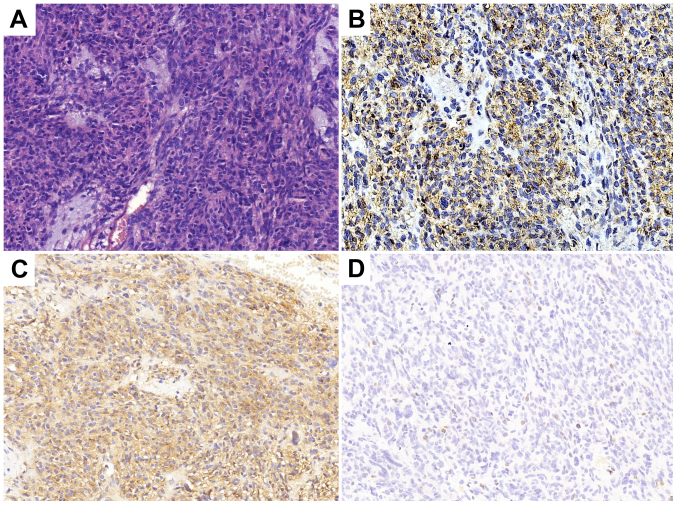
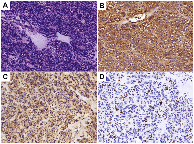
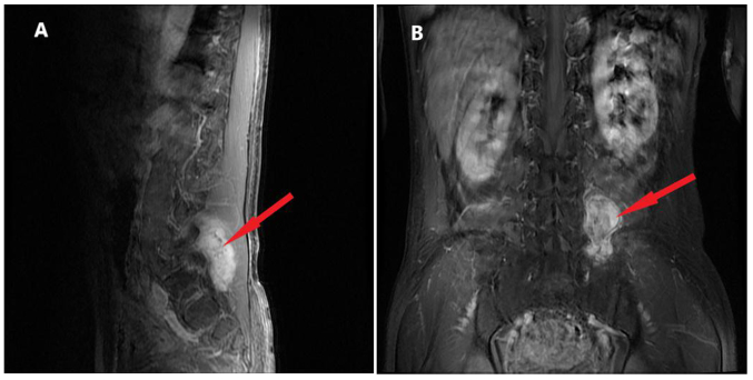
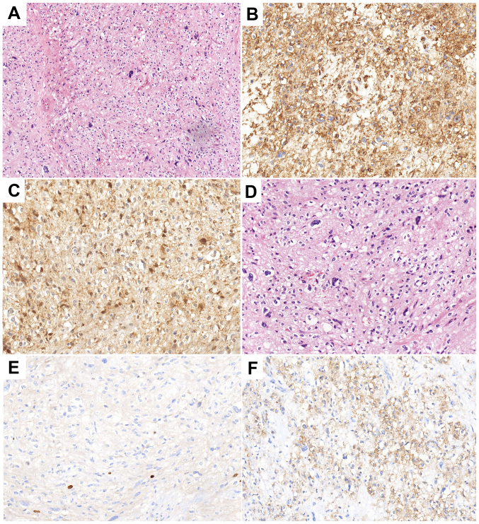
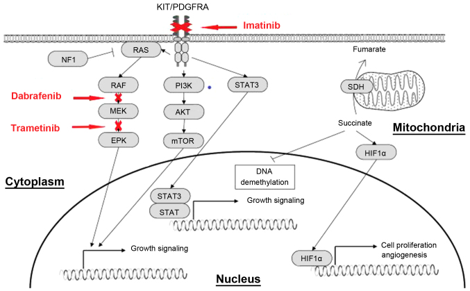
Until recently, few cases of three or more malignant tumors in one patient have been reported. Owing to the high incidence rate of these tumors, the improvement in cancer diagnosis and treatment, and the extension of patient survival time, the incidence of reported multiple primary malignant neoplasms has gradually increased. The present study reported the case of a 57-year-old man with non-small cell lung cancer combined with B-Raf proto-oncogene serine/threonine kinase V600E mutation, gastrointestinal stromal tumors and lumbar vertebral malignant mucinous sarcoma. The pathogenesis, diagnosis and treatment of these three malignancies are discussed and previous studies are also reviewed. The aim of the study was to analyze the genetic mutations associated with multiple primary malignant tumors and to discuss whether those mutations with unknown functional significance could be used as therapeutic indicators. This case report will serve as a reference for future treatment of such patients.

Keywords: BRAF V600E mutation; PD-L1; dabrafenib; gastrointestinal stromal tumor; imatinib; malignant fibromyxoid sarcoma; multiple primary malignant neoplasms; non-small cell lung cancer; trametinib.


Figures

Figure 1.
Figure 1.
Histopathological analysis following gastric lesion resection and left hepatic lobectomy in 2007. (A) Hematoxylin and eosin staining indicating the tumor as a low-grade malignant gastrointestinal stromal tumor. Tumor cells were (B) CD34 (++), (C) CD117 (+++) and (D) Ki-67 (+, <10%). Magnification, ×200.
Figure 2.
Figure 2.
Histopathological analysis following total gastrectomy, stern resection of the pancreas, splenectomy, left adrenalectomy and Roux-en-Y esophagojejunostomy in 2013. (A) Hematoxylin and eosin staining indicating a malignant gastrointestinal stromal tumor. Tumor cells were (B) CD34(+), (C) CD117(+/-) and (D) Ki-67(+, 20%). Magnification, ×200.
Figure 3.
Figure 3.
Magnetic resonance imaging of the L5 prior to the operation in 2015. (A) Sagittal and (B) coronal position showing the irregular lesion (red arrow) of 4.2×5.7×6.7 cm between the left transverse process of the L5 and sacroiliac joint, partly involving the sacrum and left iliac bone. L5, 5th lumbar vertebra.
Figure 4.
Figure 4.
Histopathological analysis results following the surgery in 2016. (A and B) Hematoxylin and eosin staining indicating a malignant ossifying fibromyxoid sarcoma with infiltrating growth and satellite nodules besides the main body of the tumor. Tumor cells were (C) CD34(+), (D) Ki67(+, 1–2%), (E) cyclin-dependent kinase 4 (+), and (F) smooth muscle actin (+). Magnification, (A) ×100 and (B and C).
Figure 5.
Figure 5.
Histopathological analysis results following cervical lymph node dissection and biopsy in 2018. (A) Hematoxylin and eosin staining indicating metastatic low-differentiated adenocarcinoma of the third and fourth group of the right cervical lymph nodes. Tumor cells were (B) transcription termination factor 1 (+) and (C) cytokeratin 7 (+). Magnification, ×200.
Figure 6.
Figure 6.
Positron emission tomography-contrast enhanced scan. (A and B) The scan of the whole body. Metabolic activity was observed in (C) the right lower lobe (red arrow), (D-F) the mediastinal lymph nodes (red arrow) and (G) the liver (red arrow). The tumor is located on (C) the inferior lobe of the right lung (red arrow), and (G) liver metastases (red arrow) was considered to be pulmonary adenocarcinoma. (H) The lymph nodes (red arrow) adjacent to the abdominal aorta
Figure 7.
Figure 7.
CT scans of foci in the lung and liver. CT scans (A-D) prior to treatment, and after (E-H) 2 and (I-L) 4 months of oral dabrafenib and trametinib treatment. CT, computed tomography. Red arrows indicate (A, E and I) the lesion in right lung and (D and H) the lesion in the liver.
Figure 8.
Figure 8.
CT scans of focus in the liver. CT scans (A) 4 and (B) 6 months after oral dabrafenib and trametinib treatment. The red arrow indicates the lesion in the liver. CT, computed tomography.
Figure 9.
Figure 9.
Effect of imatinib, dabarafenib and trametinib on downstream signaling pathways. Activating mutations of KIT and PDGFRA genes permit ligand-independent phosphorylation of the receptor tyrosine kinases, allowing the receptor-initiated signal and causing activation of the downstream effectors, such as PI3K/AKT, Ras/MAPK and JAK/STAT. PDGFRA, platelet-derived growth factor receptor α; JAK, Janus kinase; MAPK, mitogen-activated protein kinase; PI3K, phophoinositide-3 kinase; mTOR, mechanistic target of rapamycin; STAT, signal transducer and activator of transcription; c-Kit, KIT proto-oncogene receptor tyrosine kinase; HIF1α, hypoxia inducible factor-1α; SDH, succinate dehydrogenase; ERK, extracellular regulated protein kinases.

Similar articles

Cited by

References

    1. DE Luca A, Frusone F, Vergine M, Cocchiara R, LA Torre G, Ballesio L, Monti M, Amabile MI. Breast cancer and multiple primary malignant tumors: Case report and review of the literature. In vivo. 2019;33:1313–1324. doi: 10.21873/invivo.11605. - DOI - PMC - PubMed
    1. Zhao J, Tan Y, Wu Y, Zhao W, Wu J, Ji M, Shi L, Jiang J, Wu C. A rare case of eight multiple primary malignant neoplasms in a female patient: A case report and review of the literature. Oncol Lett. 2015;9:587–590. doi: 10.3892/ol.2014.2789. - DOI - PMC - PubMed
    1. Mukaiyama Y, Suzuki M, Morikawa T, Mori Y, Takeshima Y, Fujimura T, Fukuhara H, Nakagawa T, Nishimatsu H, Kume H, Homma Y. Multiple primary malignant neoplasms of the glottis, renal pelvis, urinary bladder, oral floor, prostate, and esophagus in a Japanese male patient: A case report. World J Surg Oncol. 2014;12:294. doi: 10.1186/1477-7819-12-294. - DOI - PMC - PubMed
    1. Wang H, Hou J, Zhang G, Zhang M, Li P, Yan X, Ma Z. Clinical characteristics and prognostic analysis of multiple primary malignant neoplasms in patients with lung cancer. Cancer Gene Ther. 2019 Jan 31; doi: 10.1038/s41417-019-0084-z. (Epub ahead of print) - DOI - PubMed
    1. Lee J, Park S, Kim S, Kim J, Ryu J, Park HS, Kim SI, Park BW. Characteristics and survival of breast cancer patients with multiple synchronous or metachronous primary cancers. Yonsei Med J. 2015;56:1213–1220. doi: 10.3349/ymj.2015.56.5.1213. - DOI - PMC - PubMed