Management of Bone Defects in Revision Total Knee Arthroplasty with Use of a Stepped, Porous-Coated Metaphyseal Sleeve
- PMID: 31579532
- PMCID: PMC6687494
- DOI: 10.2106/JBJS.ST.18.00038
Management of Bone Defects in Revision Total Knee Arthroplasty with Use of a Stepped, Porous-Coated Metaphyseal Sleeve
Abstract
Background: Revision total knee arthroplasty is a costly operation associated with many challenges including bone loss in the distal end of the femur and proximal end of the tibia1,2. Reconstruction of bone defects remains a difficult problem that may require more extensive reconstruction techniques to restore mechanical stability and ensure long-term fixation. Use of porous-coated metaphyseal sleeves is a modern technique to address bone deficiency in revision total knee arthroplasty3,4. Midterm reports have shown excellent survivorship and osseointegration5-7.
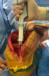
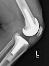
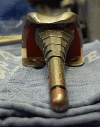
Description: The use of a porous-coated metaphyseal sleeve begins with intramedullary canal reaming to determine the diameter of the diaphyseal-engaging stem. Bone loss is assessed followed by broaching of the tibial and/or femoral metaphyses. Broaching continues until axial and rotational stability are achieved. The sleeve typically occupies most, if not all, of the proximal tibial and distal femoral cavitary osseous defects often encountered during revision total knee arthroplasty. However, a sleeve does not address all distal and posterior femoral condylar bone loss, for which augments are often required.
Alternatives: Previously described methods to address various bone deficiencies include use of morselized or structural bone-grafting, reinforcing screws within cement, metal augments, and metaphyseal cone fixation8-17.
Rationale: Structural allografts or metal augments remain a suitable option for uncontained metaphyseal defects. Metaphyseal structural allografts may undergo stress-shielding, resorption, and late fracture. Metaphyseal sleeves offer long-term biologic fixation to host bone while creating a stable platform to receive a cemented femoral and/or tibial component7. This hybrid combination may provide mechanically protective properties to decrease the loads at the cement-bone interfaces and enhance loads to metaphyseal bone to ensure long-term implant fixation in the setting of substantial bone deficiencies18-20.
Copyright © 2019 by The Journal of Bone and Joint Surgery, Incorporated.
Figures


Similar articles
-
Tibial revision knee arthroplasty with metaphyseal sleeves: The effect of stems on implant fixation and bone flexibility.PLoS One. 2017 May 8;12(5):e0177285. doi: 10.1371/journal.pone.0177285. eCollection 2017. PLoS One. 2017. PMID: 28481956 Free PMC article.
-
Porous-Coated Metaphyseal Sleeves for Severe Femoral and Tibial Bone Loss in Revision TKA.J Arthroplasty. 2017 Nov;32(11):3468-3473. doi: 10.1016/j.arth.2017.06.025. Epub 2017 Jun 20. J Arthroplasty. 2017. PMID: 28697864
-
[Knee revision arthroplasty : cementless, metaphyseal fixation with sleeves].Oper Orthop Traumatol. 2015 Feb;27(1):24-34. doi: 10.1007/s00064-014-0333-0. Epub 2015 Jan 28. Oper Orthop Traumatol. 2015. PMID: 25620192 Clinical Trial. German.
-
Techniques for filling tibiofemoral bone defects during revision total knee arthroplasty.Orthop Traumatol Surg Res. 2021 Feb;107(1S):102776. doi: 10.1016/j.otsr.2020.102776. Epub 2020 Dec 13. Orthop Traumatol Surg Res. 2021. PMID: 33321231 Review.
-
Extraction of total knee arthroplasty intramedullary stem extensions.Orthop Traumatol Surg Res. 2020 Feb;106(1S):S135-S147. doi: 10.1016/j.otsr.2019.05.025. Epub 2019 Dec 4. Orthop Traumatol Surg Res. 2020. PMID: 31812635 Review.
Cited by
-
Metaphyseal Sleeve Failure in Revision Total Knee Arthroplasty.Cureus. 2021 Sep 17;13(9):e18054. doi: 10.7759/cureus.18054. eCollection 2021 Sep. Cureus. 2021. PMID: 34692283 Free PMC article.
-
Mid-term clinical and radiographic outcomes of porous-coated metaphyseal sleeves used in revision total knee arthroplasty.Knee Surg Relat Res. 2021 May 4;33(1):16. doi: 10.1186/s43019-021-00103-5. Knee Surg Relat Res. 2021. PMID: 33947470 Free PMC article.
-
Early Survivorship of Newly Designed Highly Porous Metaphyseal Tibial Cones in Revision Total Knee Arthroplasty.Arthroplast Today. 2021 Feb 23;8:5-10. doi: 10.1016/j.artd.2021.01.004. eCollection 2021 Apr. Arthroplast Today. 2021. PMID: 33665275 Free PMC article.
-
Conversion From Knee Arthrodesis Back to Arthroplasty: A Particular Challenge in Combination With Fungal Periprosthetic Joint Infection.Arthroplast Today. 2020 Dec 5;6(4):1038-1044. doi: 10.1016/j.artd.2020.10.007. eCollection 2020 Dec. Arthroplast Today. 2020. PMID: 33385048 Free PMC article.
-
Primary total knee arthroplasty in patients with a significant bone defect in the medial tibial plateau: Case series and literature review.Int J Surg Case Rep. 2023 Sep;110:108779. doi: 10.1016/j.ijscr.2023.108779. Epub 2023 Sep 2. Int J Surg Case Rep. 2023. PMID: 37666156 Free PMC article.
References
-
- Sheth NP, Bonadio MB, Demange MK. Bone loss in revision total knee arthroplasty: evaluation and management. J Am Acad Orthop Surg. 2017. May;25(5):348-57. - PubMed
-
- Engh GA. Bone defect classification. In: Engh GA, Rorabeck CH, editors. Revision total knee arthroplasty. Baltimore: Lippincott Williams & Wilkins; 1997. p 63-120.
-
- Daines BK, Dennis DA. Management of bone defects in revision total knee arthroplasty. Instr Course Lect. 2013;62:341-8. - PubMed
-
- Dennis DA. A stepwise approach to revision total knee arthroplasty. J Arthroplasty. 2007. June;22(4)(Suppl 1):32-8. - PubMed
-
- Barnett SL, Mayer RR, Gondusky JS, Choi L, Patel JJ, Gorab RS. Use of stepped porous titanium metaphyseal sleeves for tibial defects in revision total knee arthroplasty: short term results. J Arthroplasty. 2014. June;29(6):1219-24. Epub 2013 Dec 25. - PubMed
LinkOut - more resources
Full Text Sources
