Utility of cytology in the diagnosis of parasitic infestation: A retrospective study
- PMID: 31579663
- PMCID: PMC6767801
- DOI: 10.4103/tp.TP_3_19
Utility of cytology in the diagnosis of parasitic infestation: A retrospective study
Abstract
Background: Parasitic infestation is one of the serious health problems in developing countries. Parasitic infestation is usually asymptomatic and does not cause disease as it may eventually lead to the death of both organism and host.
Materials and methods: This was a retrospective study done over a period of 5 years from 2013 to 2018. The study included 26 cases of parasitic infestations diagnosed on fine-needle aspiration cytology (FNAC) as well as fluid cytology.
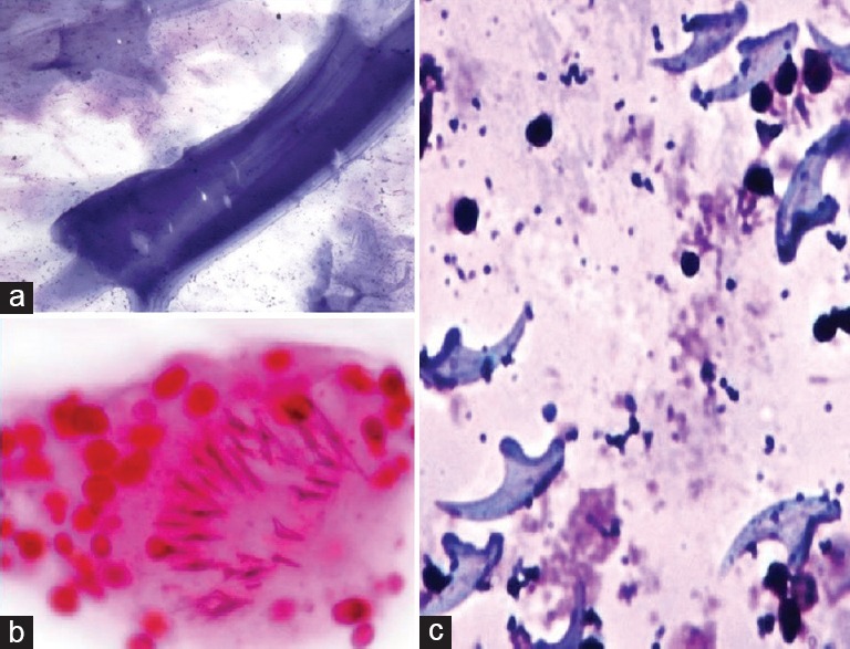
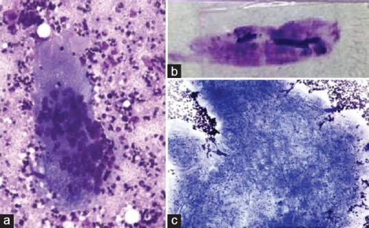
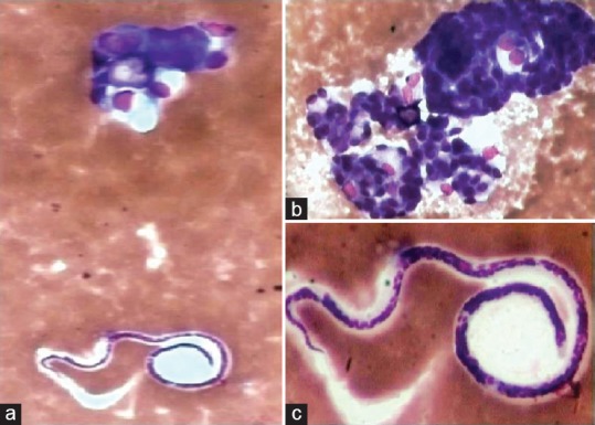
Results: Hydatidosis, cysticercosis, and filariasis were the parasitic infestations observed in this study, of which hydatidosis was the most common infestation. The predominant age group was 20-85 years old, with a mean age of presentation being 55 years. There was male predominance with a male-female of 9:1.
Conclusion: FNAC and fluid cytology are rapid diagnostic tools that aid in the early diagnosis of parasitic infestations. In parasitic infestations presenting as visceral cystic lesions, thorough examination with proper clinical correlation aid in early management. In cases with coexistent malignancy, cytology plays a major role in the diagnosis of silent carriers of infection.
Keywords: Fine-needle aspiration cytology; fluid cytology; parasitic infestation.
Copyright: © 2019 Tropical Parasitology.
Conflict of interest statement
There are no conflicts of interest.
Figures