

The role of EUS in diagnosis and treatment of liver disorders
- PMID: 31579708
- PMCID: PMC6773586
- DOI: 10.1055/a-0958-2183
The role of EUS in diagnosis and treatment of liver disorders
Abstract
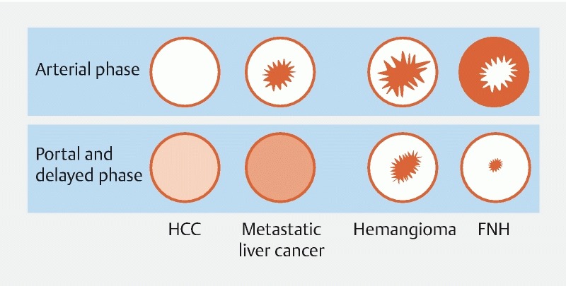
Background and aim Transabdominal ultrasound (US), computed tomographic scanning (CT) and magnetic resonance imaging (MRI) are established diagnostic tools for liver diseases. Percutaneous transhepatic cholangiography is used to perform hepatic interventional procedures including biopsy, biliary drainage procedures, and radiofrequency ablation. Despite their widespread use, these techniques have limitations. Endoscopic ultrasound (EUS), a tool that has proven useful for evaluating the mediastinum, esophagus, stomach, pancreas, and biliary tract, has an expanding role in the field of hepatology complementing the traditional investigational modalities. This review aimed to assess the current scientific evidence regarding diagnostic and therapeutic applications of EUS for hepatic diseases.
Conflict of interest statement
Figures
References
-
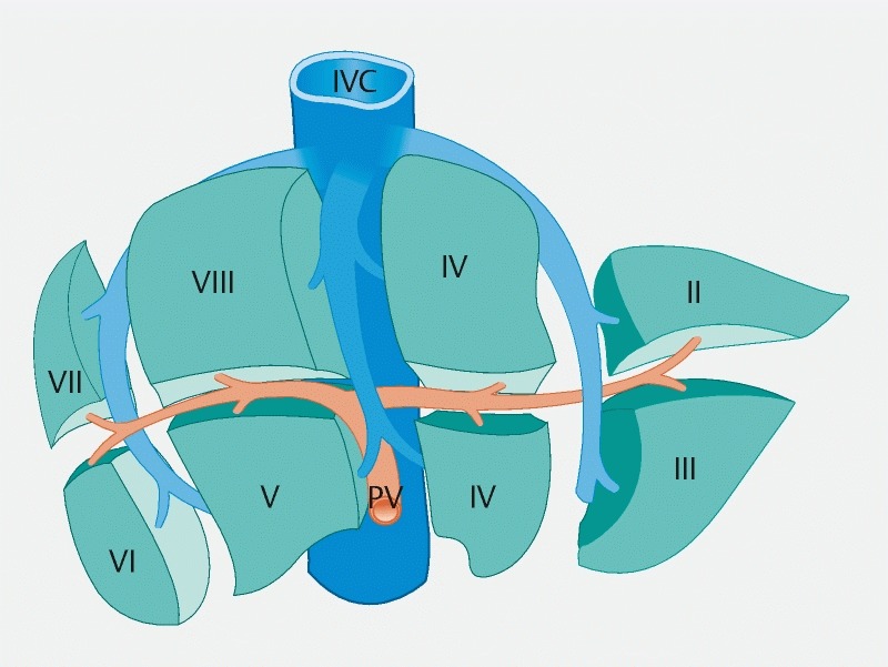
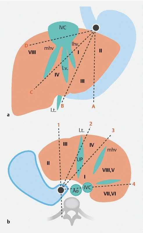
- Couinaud C. [Liver lobes and segments: notes on the anatomical architecture and surgery of the liver ] Presse Med. 1954;62:709–712. - PubMed
-
- Bhatia V, Hijioka S, Hara K et al.Endoscopic ultrasound description of liver segmentation and anatomy: Endoscopic ultrasound anatomy of liver. Dig Endosc. 2014;26:482–490. - PubMed
-
- Andanappa H K, Dai Q, Korimilli A et al.Acoustic liver biopsy using endoscopic ultrasound. Dig Dis Sci. 2008;53:1078–1083. - PubMed
-
- Singh P, Mukhopadhyay P, Bhatt B et al.Endoscopic ultrasound versus CT scan for detection of the metastases to the liver: results of a prospective comparative study. J Clin Gastroenterol. 2009;43:367–373. - PubMed
-
- Caletti G, Brocchi E, Baraldini M et al.Assessment of portal hypertension by endoscopic ultrasonography. Gastrointest Endosc. 1990;36:S21–S27. - PubMed
Publication types
LinkOut - more resources
Full Text Sources