. 2019 Sep 25;5(9):eaax2166.
doi: 10.1126/sciadv.aax2166. eCollection 2019 Sep.

Disruptive variants of CSDE1 associate with autism and interfere with neuronal development and synaptic transmission

Hui Guo  1   2 Ying Li  1 Lu Shen  1 Tianyun Wang  1   2 Xiangbin Jia  1 Lijuan Liu  3 Tao Xu  4 Mengzhu Ou  3 Kendra Hoekzema  2 Huidan Wu  1 Madelyn A Gillentine  2 Cenying Liu  1 Hailun Ni  1 Pengwei Peng  1 Rongjuan Zhao  1 Yu Zhang  5 Chanika Phornphutkul  6 Alexander P A Stegmann  7 Carlos E Prada  8 Robert J Hopkin  8 Joseph T Shieh  9 Kirsty McWalter  10 Kristin G Monaghan  10 Peter M van Hasselt  11 Koen van Gassen  11 Ting Bai  1 Min Long  1 Lin Han  1 Yingting Quan  1 Meilin Chen  1 Yaowen Zhang  1 Kuokuo Li  1 Qiumeng Zhang  1 Jieqiong Tan  1 Tengfei Zhu  1 Yaning Liu  1 Nan Pang  12 Jing Peng  12 Daryl A Scott  13   14 Seema R Lalani  13 Mahshid Azamian  13 Grazia M S Mancini  15 Darius J Adams  16 Malin Kvarnung  17   18 Anna Lindstrand  17   18 Ann Nordgren  17   18 Jonathan Pevsner  19   20 Ikeoluwa A Osei-Owusu  19   20 Corrado Romano  21 Giuseppe Calabrese  21 Ornella Galesi  21 Jozef Gecz  22 Eric Haan  23 Judith Ranells  24 Melissa Racobaldo  24 Magnus Nordenskjold  17   18 Suneeta Madan-Khetarpal  25 Jessica Sebastian  25 Susie Ball  26 Xiaobing Zou  27 Jingping Zhao  28 Zhengmao Hu  1 Fan Xia  13   29 Pengfei Liu  13   29 Jill A Rosenfeld  13 Bert B A de Vries  30 Raphael A Bernier  31 Zhi-Qing David Xu  4 Honghui Li  5 Wei Xie  3 Robert B Hufnagel  32 Evan E Eichler  2   33 Kun Xia  1   34   35   36
Affiliations

Disruptive variants of CSDE1 associate with autism and interfere with neuronal development and synaptic transmission

Hui Guo et al. Sci Adv. .

Abstract

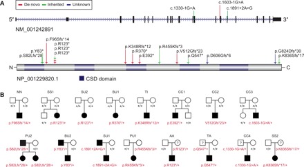
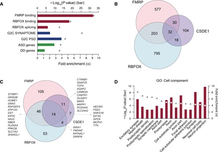
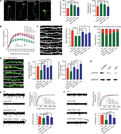
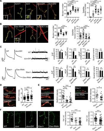
RNA binding proteins are key players in posttranscriptional regulation and have been implicated in neurodevelopmental and neuropsychiatric disorders. Here, we report a significant burden of heterozygous, likely gene-disrupting variants in CSDE1 (encoding a highly constrained RNA binding protein) among patients with autism and related neurodevelopmental disabilities. Analysis of 17 patients identifies common phenotypes including autism, intellectual disability, language and motor delay, seizures, macrocephaly, and variable ocular abnormalities. HITS-CLIP revealed that Csde1-binding targets are enriched in autism-associated gene sets, especially FMRP targets, and in neuronal development and synaptic plasticity-related pathways. Csde1 knockdown in primary mouse cortical neurons leads to an overgrowth of the neurites and abnormal dendritic spine morphology/synapse formation and impaired synaptic transmission, whereas mutant and knockdown experiments in Drosophila result in defects in synapse growth and synaptic transmission. Our study defines a new autism-related syndrome and highlights the functional role of CSDE1 in synapse development and synaptic transmission.


Figures

Fig. 1
Fig. 1. Spectrum of CSDE1 LGD variants and patient facial features.
(A) Diagram of the canonical CSDE1 isoform (NM_001242891.1 and NP_001229820.1). The locations of LGD variants are indicated. (B) Pedigrees of eight families with de novo LGD variants (above) and eight families with transmitted LGD variants (below). Carrier parents or sibling in at least four families (PU2, BU2, SU1, and PU1) are affected or show substantial family history.
Fig. 2
Fig. 2. Enrichment analyses of Csde1 RNA binding targets.
(A) Bar plot shows Csde1-binding targets significantly enriched in seven ASD-related gene sets, especially the FMRP RNA binding targets. (B) Venn diagram shows the overlap of the RNA binding targets of CSDE1 and other ASD-associated RBPs (FMRP and RBFOX). (C) Venn diagram shows the overlap of the SFARI genes that are RNA binding targets of CSDE1, FMRP, and RBFOX, with gene names indicated. (D) Bar plot shows Csde1-binding targets enriched in neuronal development– and synapse development–related cell components [Fisher’s exact test, false discovery rate (FDR)–corrected]. Top 15 significant cell components are shown.
Fig. 3
Fig. 3. Disruption of Csde1 interferes with neuronal development.
(A) Csde1 KD promoted neurite and axon growth (NC, 73 neurons; Csde1 KD1, 60 neurons; Csde1 KD2, 53 neurons; Csde1 KD1 + WT, 82 neurons). Neurons were colabeled with 4′,6-diamidino-2-phenylindole (DAPI) (nuclei), green fluorescent protein (GFP) (overall neuronal morphology), and SMI 312 (axon). Scale bar, 100 μm. (B) Csde1 KD reduced the complexity of dendritic arborization (NC, 32 neurons; Csde1 KD1, 21 neurons; Csde1 KD2, 42 neurons; Csde1 KD2 + WT, 27 neurons; Csde1 KD2 + Ctnnb1, 26 neurons). (C) Csde1 KD disrupted dendritic spine morphogenesis and maturation (NC, 27 neurons; Csde1 KD1, 41 neurons; Csde1 KD2, 23 neurons; Csde1 KD2 + WT, 40 neurons; Csde1 KD2 + Ctnnb1, 28 neurons; Csde1 KD2 + Licl, 23 neurons). Scale bar, 10 μm. (D) Csde1 KD reduced the number of excitatory (vGlut) and inhibitory (vGAT) synapses (NC, 41 neurons; Csde1 KD2, 46 neurons; Csde1 KD2 + WT, 30 neurons; Csde1 KD2 + Ctnnb1, 39 neurons; Csde1 KD2 + Licl, 28 neurons). Scale bar, 10 μm. (E and F) Voltage-clamp whole-cell recordings showed that both frequency and amplitude of mEPSCs and mIPSCs were decreased in Csde1 KD neurons (three neurons for each condition). (G) Immunoblot showed that β-catenin expression was markedly decreased in Csde1 KD neurons. Statistical data were presented as means ± SEM. *P < 0.05, **P < 0.01, ***P < 0.001, ****P < 0.0001.
Fig. 4
Fig. 4. Disruption of dUnr interferes with synapse development and transmission in Drosophila.
(A) Left: Representative NMJ4 synapses of dUnr-related lines (WT, dunr/+, dunr, dunr/Df1, and dunr/Df2). Right: Both bouton number and satellite bouton number were increased in dunr/+ line (n = 22), dunr line (n = 58), dunr/Df1 line (n = 23), and dunr/Df2 line (n = 25) compared to control (n = 61). The boutons were costained with anti-HRP labeling the neuronal plasma membrane (red) and anti-DLG (green) labeling a postsynaptic scaffold protein. Magnified image of the boxed region at left bottom shows the terminal bouton or the satellite bouton. Scale bar, 5 μm. (B) Left: Representative NMJ4 morphology of the Elav/+ line, a pan-neuronal dUnr KD line (Elav-RNAi), a Drosophila UNR rescue line (dUnr-res), and a human CSDE1 rescue line (hCSDE1-res). Right: Both bouton number and satellite bouton number were increased in Elav-RNAi line (n = 45) compared to Elav/+ line (n = 45). In dUnr-res line (n = 34) and hCSDE1-res line (n = 28), the numbers were decreased compared to Elav-RNAi line (n = 45). Scale bar, 5 μm. (C) Left: Representative traces of EJPs and mEJPs in the indicated genotypes. Right: The amplitudes of EJPs were mildly decreased in both dunr/Df1 line (WT: n = 9; dunr/Df1: n = 13) and pan-neuron KD line (Elav/+: n = 29; Elav-RNAi: n = 17). Slightly but not significantly decreased mEJP amplitude and no significant change of quantal content were observed on both KD and dunr/Df1 lines. NS, not significant. (D) The normalized fluorescent intensity of BRP was slightly increased in dunr (n = 76) and dunr/Df1 (n = 63) lines compared to controls. Scale bar, 5 μm. (E) The normalized fluorescent intensity of postsynaptic GluRIIA was markedly reduced in dunr/Df1 (n = 18), dunr/Df2 (n = 23), and KD lines (n = 25) compared to controls. Scale bar, 5 μm. (F) Decreased normalized fluorescent intensity of FM 1-43 dye was detected in dunr (n = 14), dunr/Df1 (n = 40), and KD (n = 61) lines compared to controls. Scale bar, 5 μm. Statistical data were presented as means ± SEM. *P < 0.05, **P < 0.01, and ***P < 0.001.

References

    1. Lai M.-C., Lombardo M. V., Baron-Cohen S., Autism. Lancet 383, 896–910 (2014). - PubMed
    1. American Psychiatric Association, Diagnostic and Statistical Manual of Mental Disorders, Fifth Edition (American Psychiatric Publishing, 2013).
    1. Basu S. N., Kollu R., Banerjee-Basu S., AutDB: A gene reference resource for autism research. Nucleic Acids Res. 37, D832–D836 (2009). - PMC - PubMed
    1. Sanders S. J., He X., Willsey A. J., Ercan-Sencicek A. G., Samocha K. E., Cicek A. E., Murtha M. T., Bal V. H., Bishop S. L., Dong S., Goldberg A. P., Jinlu C., Keaney J. F. III, Klei L., Mandell J. D., Moreno-De-Luca D., Poultney C. S., Robinson E. B., Smith L., Solli-Nowlan T., Su M. Y., Teran N. A., Walker M. F., Werling D. M., Beaudet A. L., Cantor R. M., Fombonne E., Geschwind D. H., Grice D. E., Lord C., Lowe J. K., Mane S. M., Martin D. M., Morrow E. M., Talkowski M. E., Sutcliffe J. S., Walsh C. A., Yu T. W.; Autism Sequencing Consortium, Ledbetter D. H., Martin C. L., Cook E. H., Buxbaum J. D., Daly M. J., Devlin B., Roeder K., State M. W., Insights into autism spectrum disorder genomic architecture and biology from 71 risk loci. Neuron 87, 1215–1233 (2015). - PMC - PubMed
    1. Iossifov I., O'Roak B. J., Sanders S. J., Ronemus M., Krumm N., Levy D., Stessman H. A., Witherspoon K. T., Vives L., Patterson K. E., Smith J. D., Paeper B., Nickerson D. A., Dea J., Dong S., Gonzalez L. E., Mandell J. D., Mane S. M., Murtha M. T., Sullivan C. A., Walker M. F., Waqar Z., Wei L., Willsey A. J., Yamrom B., Lee Y. H., Grabowska E., Dalkic E., Wang Z., Marks S., Andrews P., Leotta A., Kendall J., Hakker I., Rosenbaum J., Ma B., Rodgers L., Troge J., Narzisi G., Yoon S., Schatz M. C., Ye K., McCombie W. R., Shendure J., Eichler E. E., State M. W., Wigler M., The contribution of de novo coding mutations to autism spectrum disorder. Nature 515, 216–221 (2014). - PMC - PubMed

Publication types

MeSH terms

LinkOut - more resources