

Pseudodematiaceous Fungi in Rhinosinusal Biopsies: Report of 2 Cases With Light and Electron Microscopy Analysis
- PMID: 31579897
- PMCID: PMC6757491
- DOI: 10.1177/2632010X19874766
Pseudodematiaceous Fungi in Rhinosinusal Biopsies: Report of 2 Cases With Light and Electron Microscopy Analysis
Abstract
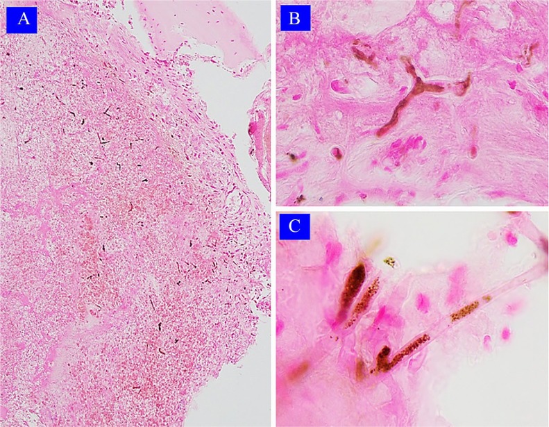
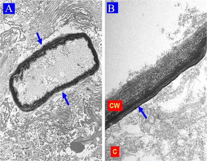
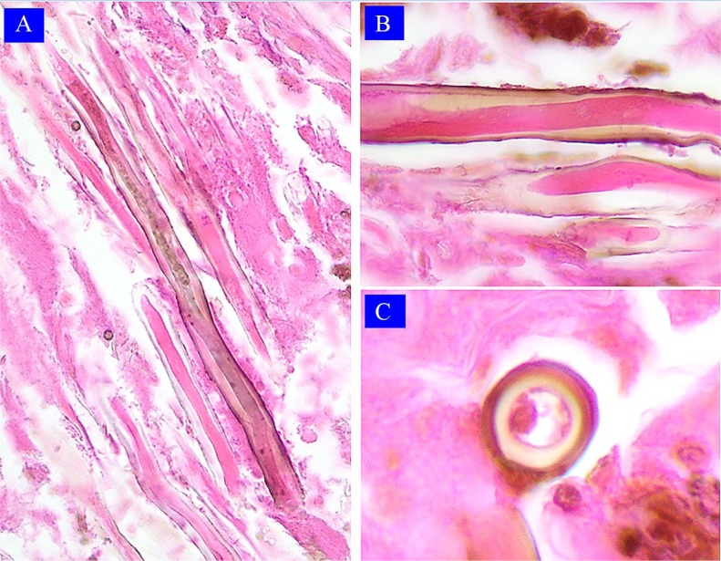
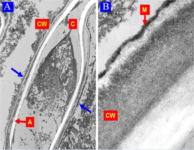
The diagnosis of a mycosis is often established through a biopsy, which allows to differentiate invasive and non-invasive lesions, and also to identify hyaline and dematiaceous fungi. However, pigmented fungal elements that do not correspond to dematiaceous fungi, which we have called pseudodematiaceous, can occasionally be present in biopsies. Herein, we present 2 cases of mycosis caused by pseudodematiaceous fungi in rhinosinusal biopsies. A new classification for fungi identified in biopsies is proposed, dividing them into 3 groups: hyaline, dematiaceous, and pseudodematiaceous.
Keywords: Pseudodematiaceous fungi; biopsy; classification; dematiaceous fungi; hyaline fungi.
© The Author(s) 2019.
Conflict of interest statement
Declaration of conflicting interests:The author(s) declared no potential conflicts of interest with respect to the research, authorship, and/or publication of this article.
Figures
Similar articles
-
Diagnostic utility of melanin production by fungi: study on tissue sections and culture smears with Masson-Fontana stain.Indian J Pathol Microbiol. 2014 Apr-Jun;57(2):217-22. doi: 10.4103/0377-4929.134666. Indian J Pathol Microbiol. 2014. PMID: 24943753
-
The Distinction between Dematiaceous Molds and Non-Dematiaceous Fungi in Clinical and Spiked Samples Treated with Hydrogen Peroxide Using Direct Fluorescence Microscopy.J Fungi (Basel). 2023 Feb 9;9(2):227. doi: 10.3390/jof9020227. J Fungi (Basel). 2023. PMID: 36836341 Free PMC article.
-
Rapid In-situ hybridization for dematiaceous fungi using a broad-spectrum oligonucleotide DNA probe.Diagn Mol Pathol. 2011 Sep;20(3):180-3. doi: 10.1097/PDM.0b013e31820e9c82. Diagn Mol Pathol. 2011. PMID: 21817900
-
Epidemiology, clinical manifestations, and therapy of infections caused by dematiaceous fungi.J Chemother. 2003 Nov;15 Suppl 2:36-47. doi: 10.1179/joc.2003.15.Supplement-2.36. J Chemother. 2003. PMID: 14708965 Review.
-
Role of In Vivo Reflectance Confocal Microscopy in the Analysis of Melanocytic Lesions.Acta Dermatovenerol Croat. 2018 Apr;26(1):64-67. Acta Dermatovenerol Croat. 2018. PMID: 29782304 Review.
Cited by
-
Pathogenesis and Pathology of COVID-Associated Mucormycosis: What Is New and Why.Curr Fungal Infect Rep. 2022;16(4):206-220. doi: 10.1007/s12281-022-00443-z. Epub 2022 Sep 29. Curr Fungal Infect Rep. 2022. PMID: 36193101 Free PMC article. Review.
-
Fungal keratitis: Mechanisms of infection and management strategies.Surv Ophthalmol. 2022 May-Jun;67(3):758-769. doi: 10.1016/j.survophthal.2021.08.002. Epub 2021 Aug 20. Surv Ophthalmol. 2022. PMID: 34425126 Free PMC article. Review.
References
-
- Sangoi AR, Rogers WM, Longacre TA, Montoya JG, Baron EJ, Banaei N. Challenges and pitfalls of morphologic identification of fungal infections in histologic and cytologic specimens. Am J Clin Pathol. 2009;131:364-375. - PubMed
-
- Nucci M, Anaissie EJ. Hyalohyphomycosis. In: Anaissie EJ, McGinnis MR, Pfaller MAS, eds. Clinical Mycology. China: Churchill Livingstone; 2009:309-328.
-
- Gómez LV, Cardona-Castro N. Phaeohyphomycosis, an emerging opportunistic fungal infection. Rev CES Med. 2016;30:66-67.
Publication types
LinkOut - more resources
Full Text Sources
