Observational Study
. 2019 Oct 2;34(10):1999-2008.
doi: 10.1093/humrep/dez139.

Tissue stiffness at the human maternal-fetal interface

Affiliations
Observational Study

Tissue stiffness at the human maternal-fetal interface

Yassen Abbas et al. Hum Reprod. .

Abstract

Study question: What is the stiffness (elastic modulus) of human nonpregnant secretory phase endometrium, first trimester decidua, and placenta?

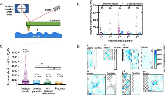
Summary answer: The stiffness of decidua basalis, the site of placental invasion, was an order of magnitude higher at 103 Pa compared to 102 Pa for decidua parietalis, nonpregnant endometrium and placenta.

What is known already: Mechanical forces have profound effects on cell behavior, regulating both cell differentiation and migration. Despite their importance, very little is known about their effects on blastocyst implantation and trophoblast migration during placental development because of the lack of mechanical characterization at the human maternal-fetal interface.

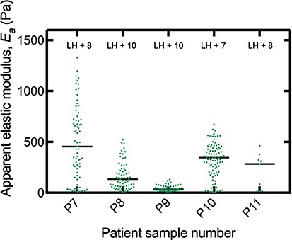
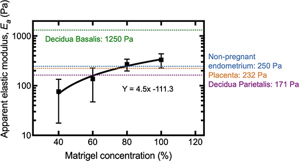
Study design, size, duration: An observational study was conducted to measure the stiffness of ex vivo samples of human nonpregnant secretory endometrium (N = 5) and first trimester decidua basalis (N = 6), decidua parietalis (N = 5), and placenta (N = 5). The stiffness of the artificial extracellular matrix (ECM), Matrigel®, commonly used to study migration of extravillous trophoblast (EVT) in three dimensions and to culture endometrial and placental organoids, was also determined (N = 5).

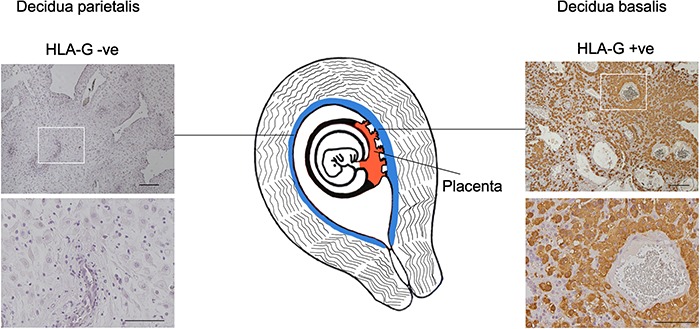
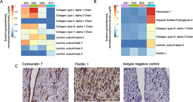
Participants/materials, setting, methods: Atomic force microscopy was used to perform ex vivo direct measurements to determine the stiffness of fresh tissue samples. Decidua was stained by immunohistochemistry (IHC) for HLA-G+ EVT to confirm whether samples were decidua basalis or decidua parietalis. Endometrium was stained with hematoxylin and eosin to confirm the presence of luminal epithelium. Single-cell RNA sequencing data were analyzed to determine expression of ECM transcripts by decidual and placental cells. Fibrillin 1, a protein identified by these data, was stained by IHC in decidua basalis.

Main results and the role of chance: We observed that decidua basalis was significantly stiffer than decidua parietalis, at 1250 and 171 Pa, respectively (P < 0.05). The stiffness of decidua parietalis was similar to nonpregnant endometrium and placental tissue (250 and 232 Pa, respectively). These findings suggest that it is the presence of invading EVT that is driving the increase in stiffness in decidua basalis. The stiffness of Matrigel® was found to be 331 Pa, significantly lower than decidua basalis (P < 0.05).

Large scale data: N/A.

Limitations, reasons for caution: Tissue stiffness was derived by ex vivo measurements on blocks of fresh tissue in the absence of blood flow. The nonpregnant endometrium samples were obtained from women undergoing treatment for infertility. These may not reflect the stiffness of endometrium from normal fertile women.

Wider implications of the findings: These results provide direct measurements of tissue stiffness during the window of implantation and first trimester of human pregnancy. They serve as a basis of future studies exploring the impact of mechanics on embryo implantation and development of the placenta. The findings provide important baseline data to inform matrix stiffness requirements when developing in vitro models of trophoblast stem cell development and migration that more closely resemble the decidua in vivo.

Study funding/competing interest(s): This work was supported by the Centre for Trophoblast Research, the Wellcome Trust (090108/Z/09/Z, 085992/Z/08/Z), the Medical Research Council (MR/P001092/1), the European Research Council (772426), an Engineering and Physical Sciences Research Council Doctoral Training Award (1354760), a UK Medical Research Council and Sackler Foundation Doctoral Training Grant (RG70550) and a Wellcome Trust Doctoral Studentship (215226/Z/19/Z).

Keywords: blastocyst implantation; human; mechanics; tissue stiffness; trophoblast invasion.


Figures

Figure 1
Figure 1
Tissues sampled at the maternal–fetal interface during first trimester pregnancy. Stiffness measurements were obtained using an AFM on decidua basalis, decidua parietalis, and placenta obtained at gestational ages between 8 and 10 weeks. Decidua was stained by IHC for HLA-G+ EVT to confirm whether the tissue was decidua basalis, while decidua parietalis was negative. Scale bars are 200 and 100 μm (insets).
Figure 2
Figure 2
AFM measurements showing increased stiffness of decidua basalis. (A) AFM was used to determine the stiffness of samples by measuring the force between a cantilever and the sample in a feedback mechanism. The reflected laser beam strikes a position-sensitive photo diode consisting of a four segment photo detector, and this is used to determine the angular deflections of the cantilever. The Hertz model can be used to estimate the apparent elastic modulus, Ea. (B) Decidua basalis was stiffer than decidua parietalis in five patient-matched samples (Mann–Whitney; ****P < 0.0001; ***P < 0.001; number of measurement points in each sample ranged from n = 10 to 148). Black bars show median for each sample. (C) A comparison of the apparent elastic modulus for decidua basalis, decidua parietalis, nonpregnant endometrium, and placenta villi; values measured for all samples are shown together. Black bars show median for each sample type (Kruskal–Wallis ANOVA with multiple comparison tests between groups; ****P < 0.001; ns means nonsignificant; N, number of patient samples; n, number of measurements). (D) Map of apparent elastic modulus values for decidua basalis and parietalis in paired samples. Each square has an area of 20 × 20μm, inside which a single measurement was taken; squares colored according to scale bar on right. Crosses represent where a clean measurement could not be taken; these are determined by evaluating the force–displacement curves manually during post-experiment analysis.
Figure 3
Figure 3
Stiffness of individual nonpregnant endometrium patient samples. Biopsies of secretory phase endometrium at 7–10 days after pre-ovulatory LH surge were tested using the AFM to determine apparent elastic modulus, Ea of each sample (N = 5 and number of measurement points in each sample ranged from n = 9 to 89). Black bars show median for each sample. Endometrium samples were stained with hematoxylin and eosin to confirm presence of luminal epithelium.
Figure 4
Figure 4
Contribution of ECM proteins to decidual stiffness. Analysis of single-cell RNA sequencing of first trimester decidua (data from Vento-Tormo et al., 2018) show normalized expression levels of RNA transcripts encoding selected ECM components. Panel (A) shows transcripts where scaled Log-transformed, normalized expression levels, LogNe in EVT formula image 1 compared with three stromal cell subsets: DS1, DS2, and DS3 identified by scRNAseq. Data from Vento-Tormo et al., 2018. Panel (B) shows ECM transcripts where expression in EVT is more that in decidual subsets, LogNeformula image1. Decidual stromal cell subsets DS1, DS2, and DS3 primarily express fibrillar collagens that provide mechanical strength. EVT cells primarily express ECM proteins that make up basement membrane. Fibrillin 1, a glycoprotein that is known to provide structural support in connective tissues, was found to be expressed by both EVT and stromal cells. (C) Protein expression of fibrillin 1 was confirmed by IHC on EVT cells. Serial sections of decidua basalis were stained for cytokeratin 7 (uterine glands and EVT) and fibrillin 1 (EVT and decidual stromal cells). Fibrillin panel shows staining of matching area to box on cytokeratin panel. White arrows indicate EVT with co-expression of both cytokeratin 7 and fibrillin 1 in serial sections (N = 3 biological replicates). Scale bars are 100 μm (cytokeratin panel) and 50μm (fibrillin panel).
Figure 5
Figure 5
An order of magnitude difference in apparent elastic modulus between Matrigel® and decidua basalis. AFM was used to determine stiffness of five batches of 100% Matrigel® (undiluted) and at dilutions to 80%, 60%, and 40% with EVT medium. Matrigel® is an artificial ECM basement membrane and the most common substrate used in 3D migration assays with EVT and culture of organoids. Nonlinear regression analysis was used to fit the data and determine the equation of the line. Matrigel® stiffness was similar to that for endometrium and first trimester placenta but much lower than that for decidua basalis.

References

    1. Abbas Y, Oefner CM, Polacheck WJ, Gardner L, Farrell L, Sharkey A, Kamm R, Moffett A, Oyen ML. A microfluidics assay to study invasion of human placental trophoblast cells. J R Soc Interface 2017;14:20170131. - PMC - PubMed
    1. Apps R, Sharkey A, Gardner L, Male V, Trotter M, Miller N, North R, Founds S, Moffett A. Genome-wide expression profile of first trimester villous and extravillous human trophoblast cells. Placenta 2011;32:33–43. Elsevier. - PMC - PubMed
    1. Bangasser BL, Shamsan GA, Chan CE, Opoku KN, Tüzel E, Schlichtmann BW, Kasim JA, Fuller BJ, McCullough BR, Rosenfeld SS et al. . Shifting the optimal stiffness for cell migration. Nat Commun 2017;8:15313 Nature Publishing Group. - PMC - PubMed
    1. Bella J, Hulmes DJS. Fibrillar collagens. Subcell Biochem 2017;82:457–490. - PubMed
    1. Benirschke K, Burton GJ, Baergen RN. Pathology of the Human Placenta, Springer. Berlin Heidelberg, 2012, http://link.springer.com/10.1007/978-3-642-23941-0. - DOI

Publication types

MeSH terms