Q-Switch Nd:YAG Laser-Assisted Decontamination of Implant Surface
- PMID: 31581536
- PMCID: PMC6960958
- DOI: 10.3390/dj7040099
Q-Switch Nd:YAG Laser-Assisted Decontamination of Implant Surface
Abstract
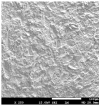
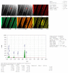
Peri-implantitis (PI) is an inflammatory disease of peri-implant tissues, it represents the most frequent complication of dental implants. Evidence revealed that microorganisms play the chief role in causing PI. The purpose of our study is to evaluate the cleaning of contaminated dental implant surfaces by means of the Q-switch Nd:YAG (Neodymium-doped Yttrium Aluminum Garnet) laser and an increase in temperature at lased implant surfaces during the cleaning process. Seventy-eight implants (titanium grade 4) were used (Euroteknika, Sallanches, France). Thirty-six sterile implants and forty-two contaminated implants were collected from failed clinical implants for different reasons, independent from the study. Thirty-six contaminated implants were partially irradiated by Q-switch Nd:YAG laser (1064 nm). Six other contaminated implants were used for temperature rise evaluation. All laser irradiations were calibrated by means of a powermetter in order to evaluate the effective delivered energy. The irradiation conditions delivered per pulse on the target were effectively: energy density per pulse of 0.597 J/cm2, pick powers density of 56 mW/cm2, 270 mW per pulse with a spot diameter of 2.4 mm, and with repetition rate of 10 Hz for pulse duration of 6 ns. Irradiation was performed during a total time of 2 s in a non-contact mode at a distance of 0.5 mm from implant surfaces. The parameters were chosen according to the results of a theoretical modeling calculation of the Nd:YAG laser fluency on implant surface. Evaluation of contaminants removal showed that the cleaning of the irradiated implant surfaces was statistically similar to those of sterile implants (p-value ≤ 0.05). SEM analysis confirmed that our parameters did not alter the lased surfaces. The increase in temperature generated at lased implant surfaces during cleaning was below 1 °C. According to our findings, Q-switch Nd:YAG laser with short pulse duration in nanoseconds is able to significantly clean contaminated implant surfaces. Irradiation parameters used in our study can be considered safe for periodontal tissue.
Keywords: biofilm; dental implant; peri-implantitis; titanium surfaces decontamination.
Conflict of interest statement
The authors declare no conflict of interest.
Figures


References
-
- Montanaro N.J., Bekov G., Romanos G.E. Optimization of thermal protocols during diode irradiation of dental implants; Proceedings of the International Laser Safety Conference; Kissimmee, FL, USA. 18–21 March 2019; p. P101. LIA.
LinkOut - more resources
Full Text Sources
Other Literature Sources
Research Materials
Miscellaneous

