. 2020 Jan;10(1):142-157.
doi: 10.1158/2159-8290.CD-19-0529. Epub 2019 Oct 3.

ID1 Mediates Escape from TGFβ Tumor Suppression in Pancreatic Cancer

Affiliations

ID1 Mediates Escape from TGFβ Tumor Suppression in Pancreatic Cancer

Yun-Han Huang et al. Cancer Discov. 2020 Jan.

Abstract

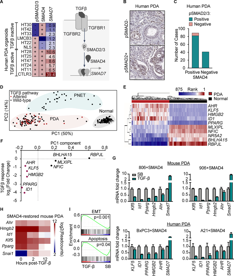
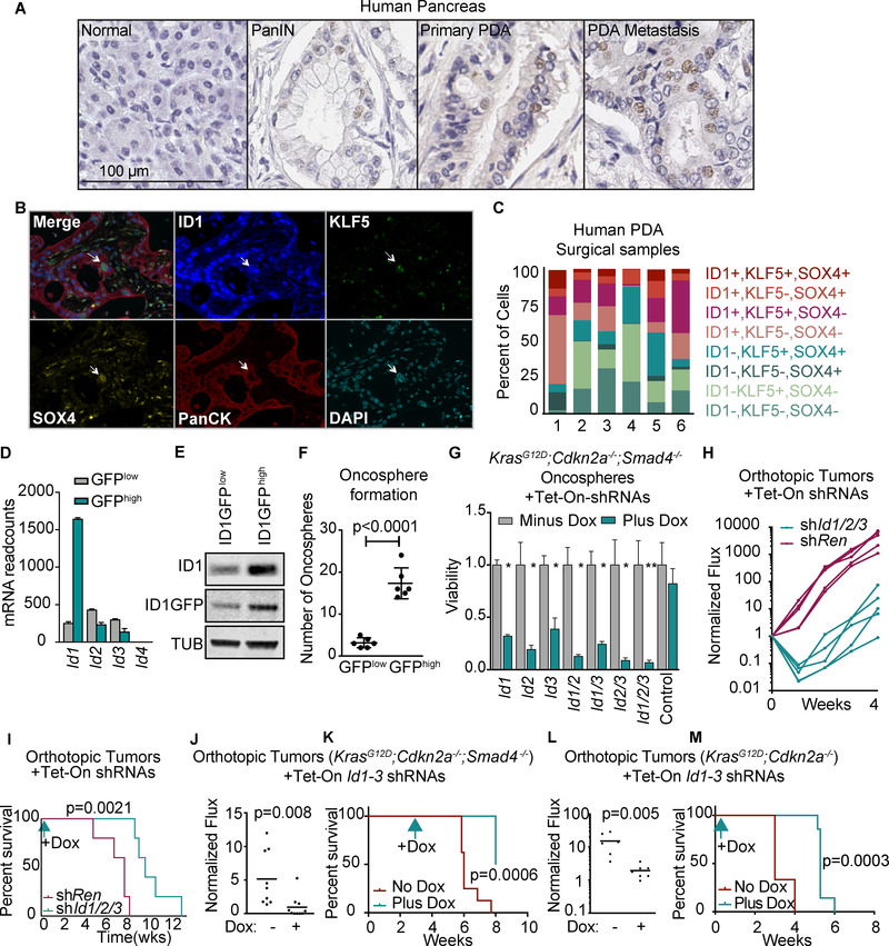
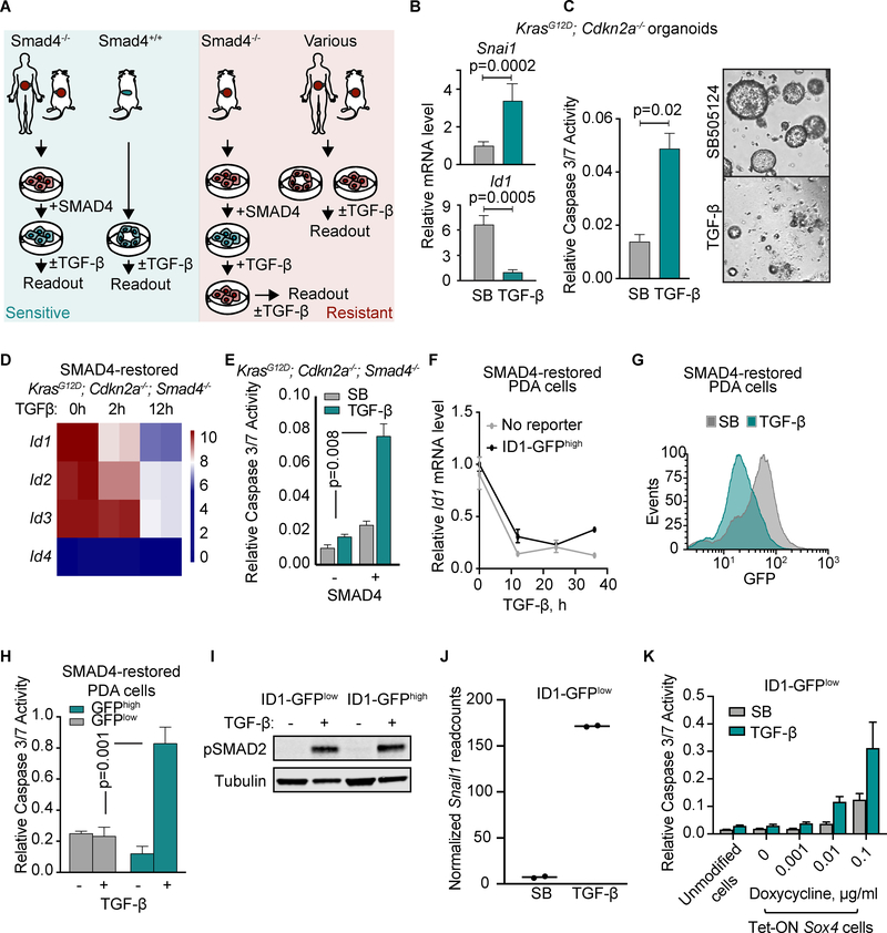
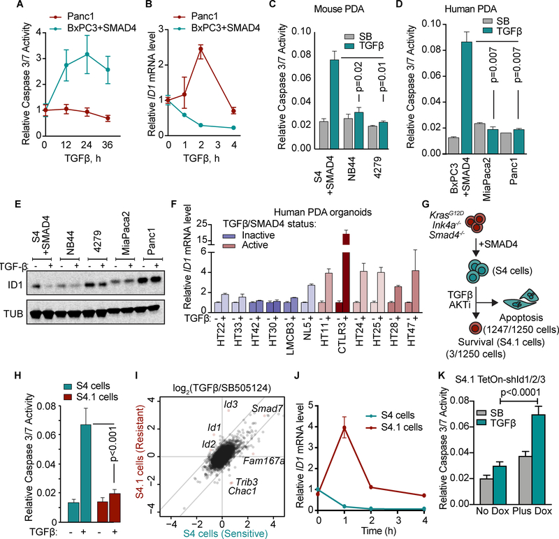
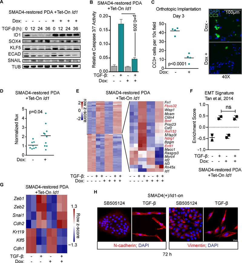
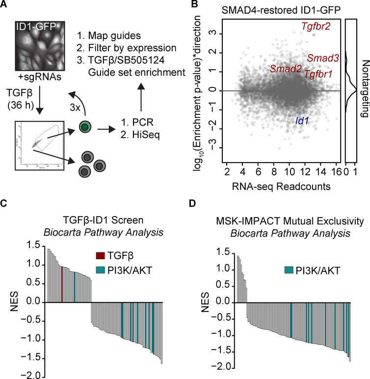
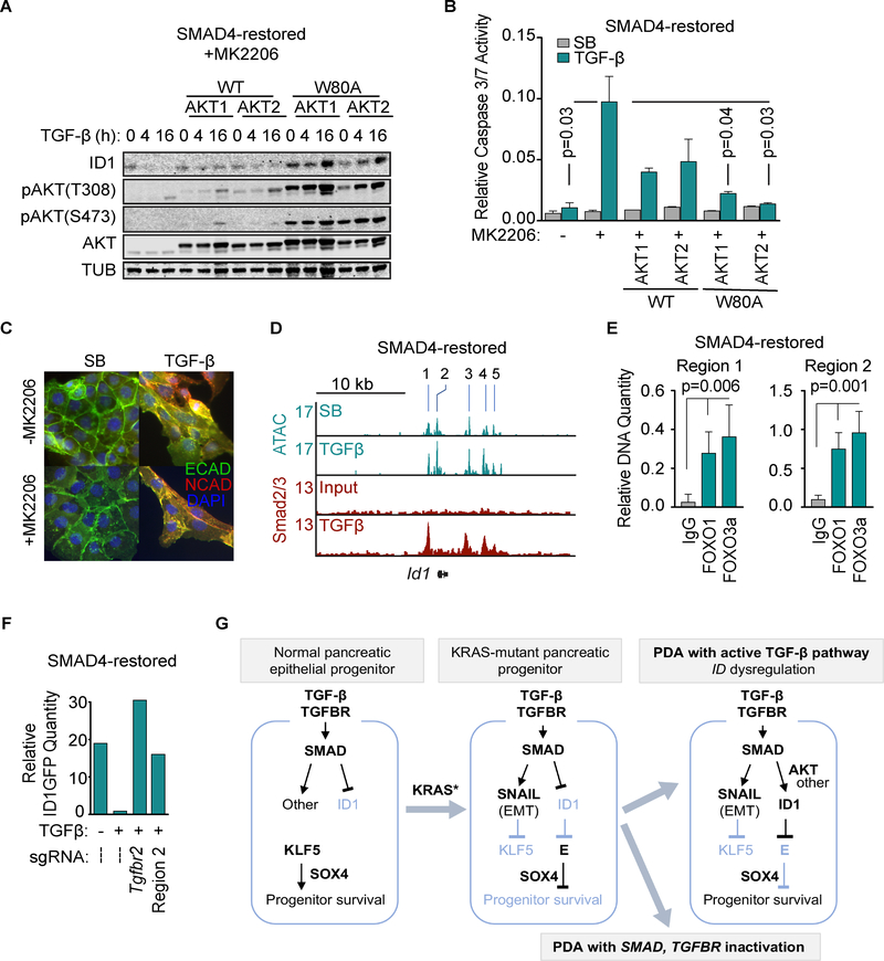
TGFβ is an important tumor suppressor in pancreatic ductal adenocarcinoma (PDA), yet inactivation of TGFβ pathway components occurs in only half of PDA cases. TGFβ cooperates with oncogenic RAS signaling to trigger epithelial-to-mesenchymal transition (EMT) in premalignant pancreatic epithelial progenitors, which is coupled to apoptosis owing to an imbalance of SOX4 and KLF5 transcription factors. We report that PDAs that develop with the TGFβ pathway intact avert this apoptotic effect via ID1. ID1 family members are expressed in PDA progenitor cells and encode components of a set of core transcriptional regulators shared by PDAs. PDA progression selects against TGFβ-mediated repression of ID1. The sustained expression of ID1 uncouples EMT from apoptosis in PDA progenitors. AKT signaling and mechanisms linked to low-frequency genetic events converge on ID1 to preserve its expression in PDA. Our results identify ID1 as a crucial node and potential therapeutic target in PDA. SIGNIFICANCE: Half of PDAs escape TGFβ-induced tumor suppression without inactivating the TGFβ pathway. We report that ID1 expression is selected for in PDAs and that ID1 uncouples TGFβ-induced EMT from apoptosis. ID1 thus emerges as a crucial regulatory node and a target of interest in PDA.This article is highlighted in the In This Issue feature, p. 1.


Figures

Figure 1:
Figure 1:. TGF-β signaling and transcriptional networks in PDA
A) Human PDA organoids were treated with or without 100 pM TGF-β for 2 h. SMAD4 and pSMAD2 were detected by Western immunoblotting (WB) and SMAD7 transcript by qRT-PCR. Values reported for SMAD7 represent fold increase induced by TGF-β. (+), strong band detected by WB; (–), weak or absent band (refer to Supplementary Fig. S1B). A schematic representation of the core TGF-β pathway components and SMAD7 as a target gene is included. B-C) A formaldehyde-fixed, paraffin-embedded tissue microarray was constructed of 130 human PDA samples collected at surgical resection and subjected to pSMAD2 and SMAD4 IHC. Samples were scored positive if ≥50% of spots contained pSMAD2 in the tumor cells (B). The number of pSMAD2+ and pSMAD2– cases in the SMAD4+ and SMAD4– groups is plotted (C). D) RNA-seq datasets of normal pancreas, PDA, and PNET from GTEx and ICGC were curated for transcription factors. Principal component analysis (PCA) was performed of the factors ranked within the top 5 of at least one case. See Supplementary Fig. S1D for complete list of factors included in the PCA. Each dot represents one tumor sample. E) Using the top 10 PC1 genes from Figure 1D, unsupervised clustering was performed of the rank-based analysis of RNA-seq datasets from GTEx and ICGC. Each column represents one tumor sample. F) SMAD4-restored KrasG12D;Cdkn2a−/−;Smad4−/− mouse PDA cells (806 cell line) were treated with 100 pM TGF-β or 2.5 μM SB505124 TGFBRI kinase inhibitor (SB) for 12 h. The samples were subjected to RNA-seq analysis. The fold-change in mRNA levels of the top 10 PC1 genes in TGF-β-treated samples compared to SB-treated samples is shown. Data are averages of two replicates per sample. G) SMAD4-restored KrasG12D;Cdkn2a−/−;Smad4−/− mouse PDA cells (806 and 906 cell lines) and SMAD4-restored human BxPC3 and A21 PDA cell lines were treated as in (F) and subjected to qRT-PCR analysis for the indicated genes H) Heatmap representation of the mRNA levels of the indicated genes in RNA-seq data sets of SMAD4-restored KrasG12D;Cdkn2a−/−;Smad4−/− mouse PDA cells treated with 100 pM TGF-β for the indicated times. Two replicates per sample. I) Gene set enrichment analysis (GSEA) of RNA-seq of SMAD4-restored mouse PDA cells treated with 100 pM TGF-β or 2.5 μM SB505124 for 12 h. Two replicates per sample.
Figure 2:
Figure 2:. ID1 expression and effects in PDA progenitors
A) Representative samples of human normal pancreas, PanIN, primary PDA, and PDA metastasis subjected to ID1 IHC analysis. B) Four-color multiplex IHC staining for ID1, KLF5, SOX4, and pan-cytokeratins in surgical PDA samples. Arrow, an ID1+,KLF5+,SOX4+ epithelial cell. C) Quantification of ID1, KLF5, and SOX4 expressing cancer cells in six surgical PDA samples (refer to Supplementary Table S1). D) Id1, Id2 and Id3 mRNA levels in sorted ID1-GFPhigh and ID1-GFPlow mouse PDA cells detected by RNA-seq. E) ID1 and ID1-GFP levels in sorted ID1-GFPhigh and ID1-GFPlow mouse PDA cells. F) KrasG12D;Cdkn2a−/−;Smad4−/− mouse PDA cells were engineered to express an ID1-GFP endogenous reporter (Supplementary Fig. S2E) and sorted based on GFP expression (Supplementary Fig. S2F). Cells were subjected to spheroid growth assays by plating 500 cells/well in low-adhesion 96-well plates, grown for 1 week, and spheroid colonies were counted. n=6 per group. Unless otherwise noted, p-values and statistics were calculated by unpaired, two-sided t-test, alpha = 0.05, as described in the Methods section. G) KrasG12D;Cdkn2a−/−;Smad4−/− mouse PDA cells were engineered with doxycycline-inducible (Tet-ON) shRNAs targeting Id genes singly or in the indicated combinations, and subjected to spheroid growth assays with or without doxycycline. Cell viability was measured by CellTiter-Glo 5 days after doxycycline addition. n=3 per group, mean ±SD, all comparisons against control. * p<0.01, ** p<0.001. H,I) KrasG12D;Cdkn2a−/−;Smad4−/− mouse PDA cells with Tet-ON shRNAs targeting Id1, Id2, and Id3 or a control shRNA (shRen) were orthotopically implanted at 500 cells per pancreas. Mice were started on doxycycline diet 3 days after implantation. (H) BLI (photon flux) was measured weekly. (I) Survival analysis. n=5 per group, p-value calculated by log-rank test. J,K) KrasG12D;Cdkn2a−/−;Smad4−/− mouse PDA cells with Tet-On shRNAs targeting Id1, Id2, and Id3 were orthotopically implanted at 500 cells per pancreas. Three weeks later, mice were randomized to matched bioluminescent groups and one group was started on doxycycline diet. n=10 per group. p-values calculated by unpaired two-sided t-test (J), and log-rank test (K). L,M) KrasG12D;Cdkn2a−/− mouse PDA cells with Tet-On shRNAs targeting Id1–3 were orthotopically implanted at 500 cells per pancreas. Three days after implantation, one group was started on doxycycline diet. Bioluminescent reading at 1 week post-implantation. n=6–7 per group, p-values calculated by unpaired two-sided t-test (L), and log-rank test (M)
Figure 3:
Figure 3:. ID1 downregulation is associated with apoptosis
A) Human and mouse PDA-derived cell models to recapitulate states that are sensitive or resistant to TGF-β-mediated apoptosis. B) qRT-PCR analysis of Id1 and Snai1 mRNA levels in KrasG12D;Cdkn2a−/− mouse organoids treated with 2.5 μM SB505124 (SB) or 100 pM TGF-β for 2h. Mean ±range of 3 replicates, representative of two independent experiments. C) Apoptosis measured by CaspaseGlo 3/7 36 h after treatment with TGF-β or SB. Two-tailed, unpaired t-test. Mean±SD of 3 replicates, representative of two independent experiments. Right, cell viability visualized by microscopy 4 days after treatment. D) Id mRNA levels from RNA-seq analysis of SMAD4-restored mouse KrasG12D;Cdkn2a−/−;Smad4−/− PDA cells treated with 100 pM TGF-β for the indicated times. E) KrasG12D;Cdkn2a−/−;Smad4−/− mouse PDA cells with and without SMAD4 restoration were treated with SB or TGF-β for 36 h. Apoptosis was measured by CaspaseGlo 3/7. Mean±SD of 3 replicates, representative of two experiments. F) qRT-PCR for Id1 in SMAD4-restored mouse PDA cells with and without the ID1-GFP reporter. Mean±range of 3 replicates, representative of two independent experiments. G) Flow cytometry analysis of GFP expression of SMAD4-restored mouse PDA cells with the ID1-GFP reporter and treated with SB or TGF-β for 36h. H) SMAD4-restored mouse PDA cells with the ID1-GFP reporter were sorted for GFP expression and then treated with 2.5 μM SB or 100 pM TGF-β for 36h. Apoptosis was measured by CaspaseGlo 3/7. Mean ±SD of 3 replicates, representative of two experiments. I) Phospho-SMAD2 levels in ID1-GFPlow and ID1-GFPhigh cells treated with SB or TGF-β for 2h. J) qRT-PCR analysis of Snai1 transcript levels in in ID1-GFPlow and ID1-GFPhigh cells treated with SB or TGF-β for 2h. K) SMAD4-restored mouse PDA cells with the ID1-GFP reporter were transduced with a Tet-On Sox4 vector, sorted for low GFP expression and treated with the indicated concentrations of doxycycline for 12h, then 2.5 μM SB or 100 pM TGF-β were added for 36h. Apoptosis was measured by CaspaseGlo 3/7. Mean ±SD of 5 replicates, representative of two experiments, two-tailed unpaired t-tests.
Figure 4:
Figure 4:. Dysregulated ID1 expression in PDAs with a functional TGF-β pathway
A,B) Human PDA cell lines with wild type SMAD4 (Panc1) or with restored SMAD4 (BxPC3+SMAD4) were treated with 100 pM TGF-β for the indicated times. Apoptosis was measured by CaspaseGlo 3/7 and ID1 mRNA levels by qRT-PCR. C) SMAD4-restored KrasG12D;Cdkn2a−/−;Smad4−/− mouse PDA cells (S4), and two KrasG12D;Cdkn2a−/− mouse PDA cell lines with wild type SMAD4 (NB44 and 4279) were treated with 2.5 μM SB505124 or 100 pM TGF-β for 36 h. Apoptosis was determined using CaspaseGlo 3/7. Mean ±SD, n=2, two-tailed unpaired t-tests. D) SMAD4-restored BxPC3 human PDA cells, and the SMAD4 wild type MiaPaca2 and Panc1 human PDA cell lines were treated with 2.5 μM SB505124 or 100 pM TGF-β for 36h and assayed using CaspaseGlo 3/7 and CellTiter-Glo. Mean±SD, n=2, two-tailed unpaired t-tests. E) S4, NB44, 4279, MiaPaca2, and Panc1 cells were treated with 2.5 μM SB505124 or 100 pM TGF-β for 24 h and subjected to ID1 and tubulin immunoblotting analysis. F) qRT-PCR analysis of ID1 mRNA levels in human PDA organoids treated with or without TGF-β for 2 h. Mean±range of 3 replicates. G) SMAD4-restored KrasG12D;Cdkn2a−/−;Smad4−/− mouse PDA cells were treated with 100 pM TGF-β and 2.5 μM MK2206 AKT inhibitor for 3 weeks, and surviving cells were selected. H) SMAD4-restored KrasG12D;Cdkn2a−/−;Smad4−/− mouse PDA cells (S4) and a resistant population selected as described in G (S4.1 cells) were treated with 2.5 μM SB or 100 pM TGF-β, in the presence of 2.5 μM MK2206, for 36 h. Apoptosis was measured using CaspaseGlo 3/7. n=6 per group, mean ±SD. I) S4 and S4.1 cells were treated with 2.5 μM SB or 100 pM TGF-β, in the presence of 2.5 μM MK2206, for 1.5h and subjected to RNA-seq analysis. Two replicates per sample. J) S4 and S4.1 cells were treated with 100 pM TGF-β for the indicated times and Id1 mRNA level was determined by qRT-PCR. Mean±range of 3 replicates, representative of 2 independent experiments. K) S4.1 cells with Tet-On shRNAs targeting Id1–3 were treated with 2.5 μM SB or 100 pM TGF-β, in the presence of 2.5 μM MK2206, for 36 h. Apoptosis was measured using CaspaseGlo 3/7. n=5 per group, mean ±SD, two-tailed unpaired t-test.
Figure 5:
Figure 5:. ID1 uncouples TGF-β-induced EMT from apoptosis
A) SMAD4-restored KrasG12D;Cdkn2a−/−;Smad4−/− mouse PDA cells transduced with a Tet-On Id1 construct were treated as indicated and subjected to immunoblotting analysis for the indicated proteins. ECAD, E-cadherin. B) The SMAD4-restored Tet-On Id1 mouse PDA cells described in (A) were incubated with or without doxycycline for 12 h and then with 2.5 μM SB505124 or 100 pM TGF-β for 36h. Apoptosis was assayed using CaspaseGlo 3/7. n=2, mean ±SD. C) The SMAD4-restored Tet-On Id1 mouse PDA cells described in (A) were treated with doxycycline for 1 day and orthotopically implanted into mice with caerulein-induced pancreatitis. Pancreata were collect at 72h for immunofluorescence analysis of cleaved caspase-3 positive (CC3+) cells. Two-tailed unpaired t-test, n=4 mice per group. D) The SMAD4-restored Tet-On Id1 mouse PDA cells described in (A) were transduced with a luciferase construct and implanted into the pancreata of mice with caerulein-induced pancreatitis. BLI was measured at 1 week. Two-tailed unpaired t-test, n=8–10 mice per group. E) The SMAD4-restored Tet-On Id1 mouse PDA cells described in (A) were treated with or without doxycycline for 12 h and then with 2.5 μM SB505124 or 100 pM TGF-β for 24h. Two replicates per sample we subjected to RNA-seq. Differentially expressed genes in response to TGF-β are represented as a heatmap on the left. The right inset shows genes that were differentially regulated ±doxycycline. F) EMT gene set variation analysis of RNA-seq dataset in E. n=2, mean±SD. G) TGF-β effects on EMT-related gene expression in the RNA-seq dataset in E. H) Cells were treated as in B for 72h and subjected to inmunofluorescence staining for the EMT mesenchymal markers N-cadherin and vimentin.
Figure 6:
Figure 6:. Mediators of ID1 dysregulation in PDA
A) Scheme of a CRISPR/Cas9 screen for mediators of ID1 dysregulation in PDA. SMAD4-restored mouse PDA cells expressing an endogenous ID1-GFP reporter and Cas9 were transduced with the Gecko_v2 genome-wide sgRNA library, selected in puromycin for 1 week, and treated for 36 h with 2.5 μM SB505124 or 100 pM TGF-β. ID1-GFPhigh cells were sorted and the process was repeated 3 times. Cells were collected for analysis of sgDNA sequences. B) sgRNAs were mapped and sgRNAs targeting genes expressed in PDA cells were selected. Gene enrichment in the TGF-β vs SB505124 samples was determined using the CAMERA competitive gene set test. Y-axis was calculated as p-value * (±1), where +1=enriched and −1=depleted. n=2 per group. C) Genes enriched in the genome-wide screen were ranked by (direction * p-value) and gene set enrichment analysis of Biocarta pathways was performed. D) MSK-IMPACT mutations mutually exclusive to TGF-β pathway alterations were analyzed via cBioPortal and ranked by –log10(p-value) for mutual exclusivity. Gene set enrichment analysis of Biocarta pathways was performed.
Figure 7:
Figure 7:. Mechanisms for loss of ID1 repression in PDA
A) SMAD4-restored mouse PDA cells were transduced with wild type or MK2206-resistant (W80A) mutant forms of AKT1 or AKT2, and treated with 2.5 μM MK2206 and 100 pM TGF-β for the indicated times. Samples were subjected to immunoblotting analysis for ID1, AKT, AKT with activating phosphorylations, or tubulin (TUB) as a loading control. B) SMAD4-restored mouse PDA cells transduced with the indicated AKT constructs were treated with or without 2.5 μM MK2206, and 2.5 μM SB505124 or 100 pM TGF-β for 36 h. Apoptosis was determined by CaspaseGlo 3/7. Two-tailed unpaired t-test, n=2 per group. C) SMAD4-restored mouse PDA cells were treated with vehicle (DMSO) or 2.5 μM MK2206 for 12h and 2.5 μM SB505124 or 100 pM TGF-β for 28h. E-cadherin and N-cadherin were detected by immunofluorescence. D) SMAD4-restored mouse PDA cells were treated with 2.5 μM SB505124 or 100 pM TGF-β for 1.5 h and subjected to ATAC-seq and SMAD2/3 ChIP-seq. Gene tracks show ATAC and SMAD2/3 ChIP tags at the Id1 locus (refer to Figure S7B for additional details) E) SMAD4-restored mouse PDA cells were treated with 2.5 μM MK2206 for 12h and then 100 pM TGF-β for 1.5h. Samples were subjected to ChIP-PCR analysis of FOXO1 and FOXO3a binding to regions 1 and 2 of the Id1 locus. Mean ±SD, two-tailed unpaired t-tests. F) SMAD4-restored mouse PDA cells expressing an endogenous ID1-GFP reporter and Cas9 were transduced with the indicated sgRNAs, treated with 2.5 μM MK2206 for 12h and then with 2.5 μM SB505124 or 100 pM TGF-β for 36h, and analyzed for GFP levels by flow cytometry. Representative of 3 independent experiments. G) Model of the role of ID1 in TGF-β tumor suppression and PDA progression. Previous work showed that KLF5 and SOX4 co-enforce of a pancreatic epithelial progenitor state and survival, whereas pancreatic progenitors harboring KRAS mutations respond to TGF-β with activation of the master EMT transcription factor SNAIL, which represses KLF5 expression. This KLF5-depleted context activates pro-apoptotic genes (3). Approximately 50% of human PDA tumors progress with inactivation of the TGF-β pathway, owing to genetic inactivation of SMAD4 and, less frequently, other core components of the TGF-β pathway (5,52). The present results identify dysregulation of ID1 as a basis for progression of the other 50% of human PDA tumors. In tumors that retain an active TGF-β pathway, activation of AKT or various low-frequency genetic alterations switch ID1 regulation by TGF-β from repression into induction. ID1 inhibits the activation of E12/E47 (E) dependent pro-apoptotic genes, thus decoupling EMT from apoptosis and allowing PDA cells to survive while retaining the ability to undergo EMT.

References

    1. David CJ, Massague J. Contextual determinants of TGFβ action in development, immunity and cancer. Nature Reviews Molecular Cell Biology 2018;164:1015–30. - PMC - PubMed
    1. Guasch G, Schober M, Pasolli HA, Conn EB, Polak L, Fuchs E. Loss of TGFbeta signaling destabilizes homeostasis and promotes squamous cell carcinomas in stratified epithelia. Cancer Cell 2007;12:313–27. - PMC - PubMed
    1. David CJ, Huang YH, Chen M, Su J, Zou Y, Bardeesy N, et al. TGF-beta Tumor Suppression through a Lethal EMT. Cell 2016;164:1015–30. - PMC - PubMed
    1. Batlle E, Massague J. Transforming Growth Factor-beta Signaling in Immunity and Cancer. Immunity 2019;50:924–40. - PMC - PubMed
    1. TCGA. Integrated Genomic Characterization of Pancreatic Ductal Adenocarcinoma. Cancer Cell 2017;32:185–203 e13. - PMC - PubMed

Publication types

MeSH terms

Substances