. 2019 Oct 3;6(6):e622.
doi: 10.1212/NXI.0000000000000622. Print 2019 Nov.

Vitamin D enhances responses to interferon-β in MS

Affiliations

Vitamin D enhances responses to interferon-β in MS

Xuan Feng et al. Neurol Neuroimmunol Neuroinflamm. .

Abstract

Objective: To determine the effect of vitamin D3 on interferon-β (IFN-β) response and immune regulation in MS mononuclear cells (MNCs).

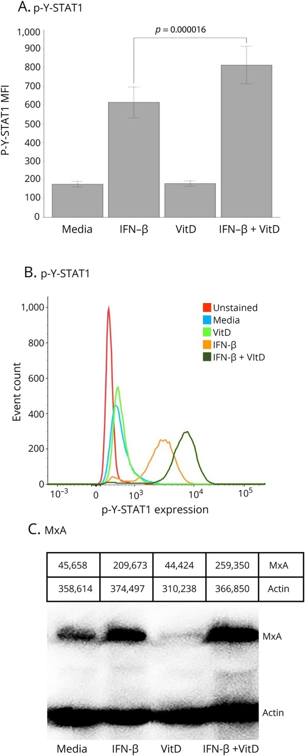
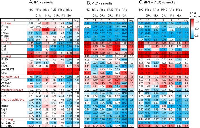
Methods: MNCs from 126 subjects, including therapy-naive patients with different forms of MS, plus patients with MS receiving IFN-β or glatiramer treatment, plus healthy controls were incubated in vitro with IFN-β-1b ± vitamin D3 (calcitriol). Activation of the IFN-β-induced transcription factor, p-Y-STAT1, and antiviral myxovirus A (MxA) protein was measured with flow cytometry and Western blots; serum proteins were measured with a customized 31-protein multiplex assay.

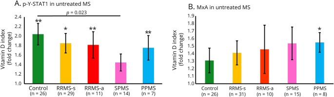
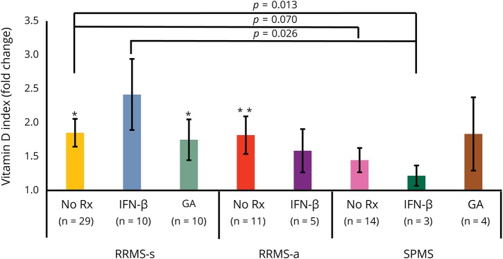
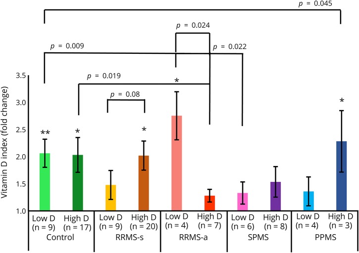
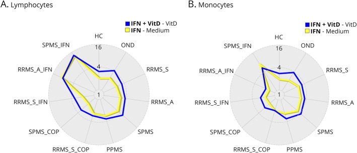
Results: Vitamin D enhanced in vitro IFN responses, as measured by induction of p-Y-STAT1 and MxA in MNCs, T cells, and monocytes. Vitamin D augmentation of IFN responses was seen in untreated and in IFN-β-1b-treated MS. The combination of vitamin D plus IFN-β reduced Th1 and Th17 cytokines, and increased Th2 responses, reversing the effect of IFN-β alone. Exacerbations and progression in untreated patients reduced the vitamin D enhancement of IFN responses. Vitamin D had less effect on IFN response in clinically stable glatiramer-treated than in IFN-β-treated patients.

Conclusion: Vitamin D enhances IFN-β induction of multiple proteins and also reverses the Th1/Th2 bias in MS seen with IFN-β alone. The combination of vitamin D and IFN-β has potential benefit in ameliorating MS.


Figures

Figure 1
Figure 1. Vitamin D enhances IFN-β–induced expression of p-Y-STAT1 and MxA in MNCs
(A) In vitro vitamin D enhances IFN-β induction of p-Y-STAT1 in ConA-activated MNCs from 72 untreated patients with MS (p = 0.00002). (B) p-Y-STAT1 expression on flow cytometry histograms from a representative patient with RRMS-s. The media, IFN, VitD, and IFN+VitD curves are all stained with Alexa Fluor 488–labeled Mab to p-Y-STAT1. (C) MxA protein on Western blot of a representative patient with RRMS-s. ConA-activated MNCs were incubated in vitro with 160 U/mL IFN-β-1b for 30 minutes to 48 hours ± preincubation with 200 nM Vit D3 (calcitriol) for 12 hours. Gray-scale densities of each band are listed above figure. IFN-β = interferon-β; MxA = myxovirus protein.
Figure 2
Figure 2. Vitamin D enhances intracellular IFN-β responses in therapy-naive MS and HCs
(A) p-Y-STAT1, measured with flow cytometry, is generated in all groups, but there is less induction in SPMS than in HC. (B) MxA protein, measured with Western blots. The trend for enhanced responses in PPMS vs HC was not significant (p < 0.13, unpaired t test). ConA-activated MNCs were incubated in vitro with 160 U/mL IFN-β-1b for 30 minutes to 48 hours ± preincubation with 200 nM vitamin D3 (calcitriol) for 12 hours. Fold change of vitamin D plus IFN-β compared with IFN-β alone, determined using VDSI: [(IFN + VitD) − (VitD)]/[(IFN) − (no IFN)]; average with p values above SEM bar: *p < 0.05, **p < 0.01. Comparisons between groups use unpaired t tests, brackets. PPMS = primary progressive MS; RRMS-a = active relapsing/remitting MS; RRMS-s = stable RRMS; SPMS = secondary progressive MS.
Figure 3
Figure 3. Vitamin D enhances IFN-β–induced p-Y-STAT1 expression in MNCs from patients with MS receiving MS therapies
In all groups, there is enhanced induction of p-Y-STAT1, measured with flow cytometry. Methods and calculations as in figure 2. *p < 0.05, **p < 0.01. IFN-β = interferon-β; RRMS-a = active relapsing/remitting MS; RRMS-s = stable RRMS; SPMS = secondary progressive MS.
Figure 4
Figure 4. Effect of high vs low serum vitamin D levels from therapy-naive patients with MS on IFN response in MNCs in vitro
Vitamin D plus IFN-β–induced p-Y-STAT1 expression is quantitated with flow cytometry. The median vitamin D level for the entire cohort was 30; low < 30, high ≥ 30 ng/mL 25-OH vitamin D. Methods and calculations as in figure 2. *p < 0.05, **p < 0.01. RRMS-a = active relapsing/remitting MS; RRMS-s = stable RRMS; PPMS = primary progressive MS; SPMS = secondary progressive MS.
Figure 5
Figure 5. Vitamin D and IFN-β effects on p-Y-STAT1 induction in lymphocytes (A) and monocytes (B)
Vitamin D plus IFN-β (blue line) in most therapy-naive groups had a greater effect than IFN-β alone (yellow line) on activation of STAT1 in ConA-activated lymphocytes and monocytes. Values in radar plot represent median fold change in log2 scale expression of p-Y-STAT1 measured by flow cytometry. Blue line: (IFN+ VitD) − (Vit D); yellow: (IFN)-(media alone). IFN-β = interferon-β; RRMS-a = active relapsing/remitting MS; RRMS-s = stable RRMS; SPMS = secondary progressive MS.
Figure 6
Figure 6. Multiplex of fold change of protein secretion by activated MNCs from HCs and different forms of MS
(A) IFN-β alone vs media, expressed as ratio of IFN-β/media. (B) Vitamin D alone vs media. (C) IFN-β plus vitamin D vs media alone. Selected targets are grouped into Th1, Th17, and Th2 immune pathways and clusters of IFN-stimulated proteins (ISGs), adhesion molecules, and cytokines controlling homeostatic proliferation, neurotrophic factors, and IL-12 family. Values are fold change in the stimulation condition/media. Intensity of shading shows upregulation (red) or downregulation (blue). Target proteins include BDNF = brain-derived neurotrophic factor; HGF = hepatocyte growth factor; IFN-γ = interferon-γ; IL-2 = interleukin-2; TNF-α = tumor necrosis factor-α; TNFRII = TNF receptor type II; IL-17F, IL-4, IL-5, IL-10, IP-10 = IFN-induced protein-10 (CXCL10); MCP1 = macrophage chemotactic protein-1 (CCL2); I-TAC = IFN-inducible T-cell alpha chemoattractant (CXCL11); LIF = leukemia inhibitory factor; p-Y-STAT1 = phosphotyrosine-STAT1 transcription factor; MxA = myxovirus A; NGF = nerve growth factor; sICAM-1 = soluble intercellular adhesion molecule-1; TPO = thymopoietin; VCAM-1 = vascular cell adhesion molecule; VEGF-α = vascular endothelial growth factor-α, IL-15, IL-7, and IL-12 p40 and IL-12 p70 components.

References

    1. Sintzel MB, Rametta M, Reder AT. Vitamin D and multiple sclerosis: a comprehensive review. Neurol Ther 2018;7:59–85. - PMC - PubMed
    1. Orton SM, Wald L, Confavreux C, et al. Association of UV radiation with multiple sclerosis prevalence and sex ratio in France. Neurology 2011;76:425–431. - PMC - PubMed
    1. Munger KL, Levin LI, Hollis BW, Howard NS, Ascherio A. Serum 25-hydroxyvitamin D levels and risk of multiple sclerosis. JAMA 2006;296:2832–2838. - PubMed
    1. Ascherio A, Munger KL, White R, et al. Vitamin D as an early predictor of multiple sclerosis activity and progression. JAMA Neurol 2014;71:306–314. - PMC - PubMed
    1. Soilu-Hänninen M, Aivo J, Lindström BM, et al. A randomised, double blind, placebo controlled trial with vitamin D3 as an add on treatment to interferon β-1b in patients with multiple sclerosis. J Neurol Neurosurg Psychiatry 2012;83:565–571. - PubMed

Publication types